
Williams Cancer Institute +1 (863) 451-9755
Lymphoma

Lymphoma, treated through one node a needle can reach.

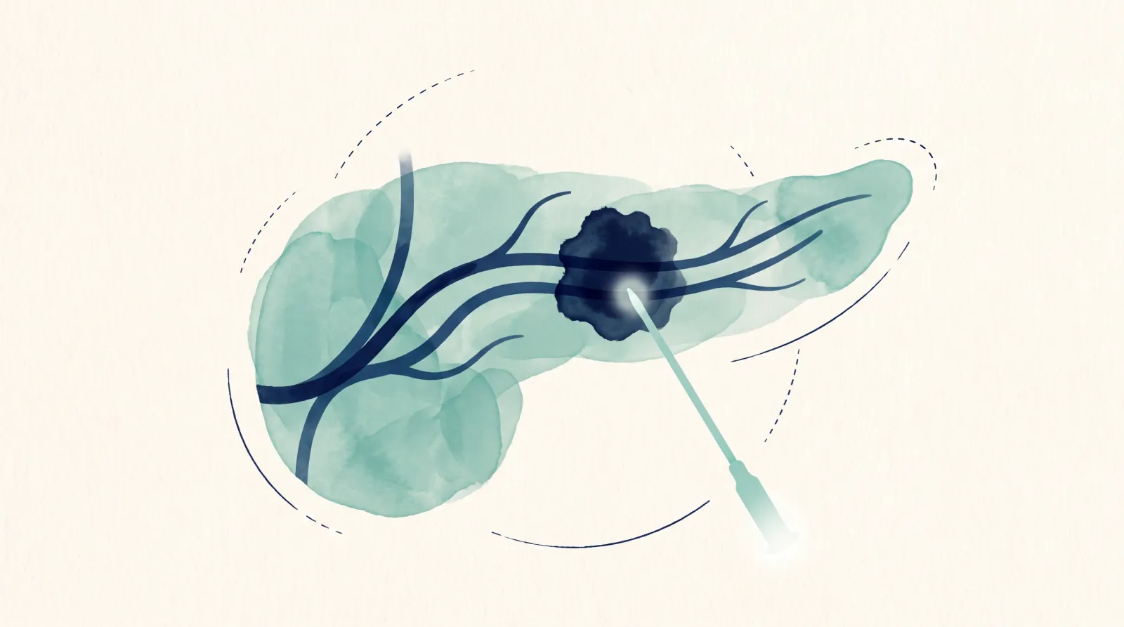
Lymph nodes are easy to reach with a needle. We treat one accessible node directly and inject immunotherapy into it, to re-engage your immune system against the rest. A physician reads your scans first, free. For lymphoma that has come back after even the newest treatments.

Start Your Admissions Process How It Works

Complete a short, three-step form to begin your admissions process. One of our patient consultants will contact you to explain the process and guide you through the next steps.

What we do
AblationVaxTreat the tumor. Teach the immune system. Treat locally, act globally.Everything we do is built on AblationVax, the approach Dr. Williams developed: the tumor is treated through a needle (ablation), and the treated tumor becomes an in-situ vaccine: a vaccine made inside your body, from your own cancer.

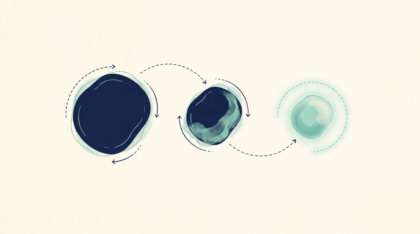
One node treated, to re-engage your immune system

CAR-T cells (lab-altered immune cells) and bispecific antibodies (immune-linking drugs) each hunt one marker on the cancer. When the cancer stops showing that marker, they stop working. We treat one node by needle and inject immune drugs there, so your immune system recognizes the cancer's other markers. It works alongside your current treatment, not in place of it.

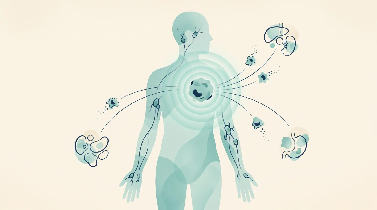
The immune drugs placed in the tumor teach the immune system to recognize this cancer, the way a vaccine does. You teach the immune system in one spot, and it goes after the same cancer in the rest of the body.

1Treat one nodeA needle reaches one lymph node and treats it there, releasing the cancer's markers.
2Treat it in placeImmune drugs go into that exact spot, in the same session.
3Teach the whole bodyThe immune system learns the cancer at one tumor and goes after it everywhere else. Doctors call this the abscopal effect. It does not stop there: the cycle below keeps it going.
Video (1:22): Why do electricity and immunotherapy work so well together? Dr. Williams explains AblationVax.
After step 3: how AblationVax keeps working

The intratumoral immune conversion cycle

The treated tumor is the source of the vaccine, and it keeps supplying it. It is not one exposure and done. Every round of tumor cells the immune system destroys exposes new antigens, and those restart the cycle, so the response keeps spreading: broader, throughout the body, and with memory. It turns a “cold” tumor, one the immune system ignores, “hot”.

01DisruptAblation breaks the tumor open and exposes its antigens, the markers the immune system uses to recognize the cancer: not a few, all of them.
02Prime and recruitImmune drugs in that same spot call immune cells in and show them the cancer.
03Activate and co-stimulateCo-stimulation is a second go-signal. Those immune cells are switched on against the antigens the ablation just exposed.
04ReleaseThe checkpoint drugs we inject take the brakes off, so the immune system can kill tumor cells.
05DestroyImmune cells destroy tumor cells, and every cell they kill exposes new antigens.
06RepeatThe new antigens restart the cycle: re-priming, amplifying and spreading the response.

Card 06 leads back to card 02. Doctors call the widening response epitope spreading.

Most cancer vaccines: outside-inMade outside the body from a few pre-selected antigens, the same for everyone, then injected. The response is limited by the antigens someone chose.
AblationVax: inside-outMade inside your body, from every antigen in your own tumor. A vaccine made for one person: you. Not a patch: a rebuilding of the immune system from within.

Lymph nodes are easy to reach

Most lymphoma sits in lymph nodes or masses a needle can reach under ultrasound or CT. That makes it one of the most accessible cancers for a direct, image-guided procedure.
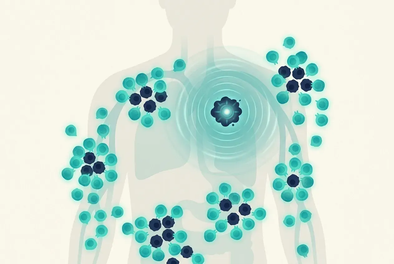
When the cancer hides the marker, we look for the others

CAR-T cells and bispecific antibodies fail when the cancer drops the CD19 or CD20 marker. Injecting immune drugs into a node is being studied as a way to re-engage your immune system against the cancer's other markers.
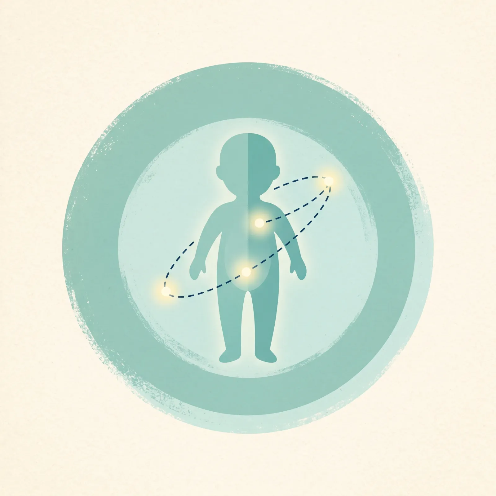
A short procedure, not an operation

About an hour under image guidance, and most people go home the same day, about two hours later.

A response across the disease

Injecting immune stimulants into one lymphoma node has research behind it, called in-situ vaccination (vaccinating at the tumor itself), mainly in slower-growing lymphomas. The immune response raised at one node reaches the disease elsewhere in the body. The cycle above is how that response keeps renewing itself.

Standard care. First-line treatment is chemotherapy-based: R-CHOP or Pola-R-CHP for large B-cell lymphoma, ABVD for Hodgkin lymphoma, bendamustine-rituximab for indolent disease. At first relapse the options are salvage chemotherapy with autologous stem-cell transplant, CAR-T cells, or CD20×CD3 bispecific antibodies. In Hodgkin lymphoma, intravenous checkpoint blockade is effective but of limited durability; brentuximab is also used. There is no established option after both CAR-T and a bispecific antibody have failed.

Why those treatments fail. CAR-T cells and bispecific antibodies act on one marker on the cancer cell (CD19 or CD20); brentuximab acts on CD30. When the lymphoma stops displaying that marker (antigen escape), or the cell product is exhausted, the treatment stops working.

What is done here. Image-guided ablation of one accessible lymph node or mass, followed in the same session by injection of a combination of immune-active agents into it. Lymphoma is unusually accessible to this: most disease sits in nodes or masses a needle can reach under ultrasound or CT. The rationale is in-situ vaccination: ablating one node exposes the lymphoma's other antigens, and the injected agents are intended to engage the patient's own immune cells against them, independent of the marker the earlier product targeted. This combination is offered under physician review. In our clinical observations, treating one node produces immune activity across the disease.

The procedure, step by step

  1. Case review. PET-CT and biopsy reports, including subtype, CD19 / CD20 / CD30 status and MYC / BCL2 / BCL6 status, are read by the Williams Cancer Institute team.
  2. Protocol design. The intratumoral combination is tuned to lymphoma subtype, prior CAR-T or bispecific therapy, and immune profile.
  3. Guidance and ablation. Image-guided percutaneous access to an accessible nodal or extranodal site.
  4. Intratumoral injection. Through the same access, into the ablation bed, in the same session.
  5. Setting and time. Most procedures are outpatient under conscious sedation; general anesthesia or an overnight stay is rarely required. About an hour for the procedure and about two hours' recovery.
  6. Follow-up. PET-CT at 6 to 8 weeks. Repeat procedures as indicated; the number of treatments is decided with the patient's physician. The patient's hematology, transplant and cellular-therapy teams are kept informed.

Agents. The injected immunotherapy is a multi-agent combination. The agents and doses are set per patient by the treating physician.

The immune conversion cycle, in clinical terms. Ablation disrupts the tumor and exposes its full antigen repertoire, not a pre-selected subset. The injected immunotherapy then remodels the tumor microenvironment, priming, recruiting and activating immune cells and releasing the brakes on them. Immune-mediated lysis exposes further antigens, which re-prime the response (epitope spreading), producing a broader, polyclonal, systemic response with memory, converting a cold tumor hot. This is WCI’s stated rationale, in Dr. Goodyear’s framing, borne out in our clinical observations.

Evidence. The pharmacological case for the intratumoral route comes from a mouse study: in mice with colorectal tumors and no ablation, anti-CTLA-4 injected into a tumor reached about 14 times the day-7 level of intravenous dosing in that tumor, the untreated tumor received no more than with intravenous dosing, and half the dose reached the blood within an hour [1]. That is mouse data in colorectal cancer; it does not test ablation and does not support a distant-site effect.

In-situ vaccination has a research precedent in lymphoma: the Stanford program studied intratumoral CpG (a TLR9 agonist) with low-dose radiation in low-grade B-cell lymphoma. In the 2025 phase I report, adding anti-OX40 (given intratumorally and intravenously) to intratumoral SD-101 plus low-dose radiation produced 1 partial response and 9 stable disease among 14 patients, inferior to the group's earlier experience with the TLR9 agonist plus radiation alone [2]. No published WCI lymphoma series exists. The combination of ablation plus multi-agent intratumoral immunotherapy in post-CAR-T or post-bispecific lymphoma has no trial behind it. Sources

  1. Meyblum L, et al. Journal for ImmunoTherapy of Cancer 2023. PMID 37949616. DOI 10.1136/jitc-2023-007433.
  2. Shree T, et al. Clinical Cancer Research 2025. PMID 39745391.
Is this for you?

Where this fits, stage by stage

Where you are in treatment decides where we fit, and for lymphoma that is almost always after the standard lines have been tried. Pick the card that sounds like your situation. Whether this is right for you is decided case by case by a physician reading your scans and pathology, so none of these lists is a verdict.

First treatment

Chemotherapy-based combinations are the standard and carry the strongest evidence (your team may call them R-CHOP, ABVD or similar). That comes first; in selected cases we work alongside your team.

First relapse

Standard care is salvage chemotherapy with a stem-cell transplant, CAR-T cells, or a bispecific antibody. These come first; in selected cases we work alongside them.

After CAR-T or a bispecific

The cell therapy or antibody has stopped working, often because the cancer dropped the marker it targeted. Options narrow here. We treat one accessible node directly to engage your own immune cells, independent of the earlier product.

A few growing spots on treatment

Most of the disease is controlled, but one or two sites are growing. Those sites can be treated directly while the immune system is engaged.

You may be a candidate if:

Any of these describes you:

  • Your large B-cell or follicular lymphoma has come back after CAR-T or a bispecific antibody
  • Your Hodgkin lymphoma has come back after checkpoint immunotherapy (drugs such as nivolumab or pembrolizumab) and brentuximab (Adcetris)
  • Your cancer has stopped showing the marker the last treatment targeted (your report may say CD19, CD20 or CD30)
  • A few spots are growing while the rest is controlled
  • You want to add immune treatment to the plan you are on
  • You are looking for options beyond your home country

It may not be suitable if:

  • You are still recovering from serious CAR-T side effects (the ones your team calls CRS or ICANS)
  • You have a serious infection, or a bleeding problem that cannot be corrected first
  • Your bone marrow is failing without a plan to support it
  • You are very unwell

Each case is individually evaluated by the Williams Cancer Institute team.

  • First line. Standard care: R-CHOP, Pola-R-CHP, ABVD, or bendamustine-rituximab for indolent disease, which carry the strongest evidence. This approach is rarely used at this step.
  • First relapse. Standard care: salvage chemotherapy with autologous transplant, CAR-T cells, or bispecific antibodies; these come first. This approach is rarely used at this step.
  • After CAR-T or a bispecific antibody. Options narrow after progression. Added here, under physician review: ablation of one node plus intratumoral immune amplification, intended to engage the patient's own T-cell response independent of the earlier product.
  • Oligoprogressive disease. A few growing sites on systemic therapy. Added here: direct treatment of those sites with intratumoral immune amplification.

WCI's stated candidacy criteria. Relapsed or refractory diffuse large B-cell lymphoma after CAR-T · progression after a CD20×CD3 bispecific antibody · follicular lymphoma after CAR-T or a bispecific · Hodgkin lymphoma after checkpoint blockade and brentuximab · antigen-escape disease (loss of CD19, CD20 or CD30).

WCI's stated exclusions. Cytokine release syndrome or neurotoxicity (CRS / ICANS) still resolving · active severe infection · uncorrectable bleeding disorder · very poor performance status (ECOG 3 or worse) · bone marrow failure without a supportive plan. Each case is evaluated individually by the Williams Cancer Institute team.

How it works

Your Admissions Process, in Five Steps

After completing our admissions form, one of our Patient Consultants will contact you to explain the process and guide you through the next steps. Our admissions process is free of charge, including the review of your medical information and, if our medical team determines that you are a candidate, a video consultation. Treatments are generally not covered by health insurance, and WCI does not bill insurance companies.

  1. 01Free

    Initial Contact

    One of our Patient Consultants will contact you to explain the admissions process, answer your general questions, and guide you through the next steps.

  2. 02Free

    Medical Records and Imaging

    Your Patient Consultant will guide you on the required medical records and imaging studies, as well as how to submit them so our medical team can evaluate your case.

  3. 03Free

    Tumor Board Review

    Once the necessary information has been received, our medical team will review your case to determine whether you may be a candidate for our treatments. If necessary, the medical team may request additional studies or information before making a decision.

  4. 04Free

    Video Consultation

    If our medical team determines that you are a candidate, we will coordinate a complimentary Zoom consultation with one of our physicians. During this consultation, you will be able to discuss your case, address your medical questions, and learn about the treatment approach indicated for you.

  5. 05Self-pay treatment

    Treatment Plan and Next Steps

    Following your medical consultation, you will receive a personalized treatment plan that includes the corresponding costs. If you decide to proceed, the next steps will be coordinated to schedule your treatment.

    Most treatments take place in Los Cabos, Mexico; in some cases our medical team may schedule a procedure in Mexico City. Rebyota is given only at our Beverly Hills, California location.

For step 05

What to expect

Your physician confirms the details for your case.

Procedure
About an hour. Most people go home the same day.
Anesthesia or a stay
Conscious sedation is the predominant anesthesia used. It is rare that procedures require general anesthesia or an overnight stay. Your physician tells you which.
Number of treatments
Determined during the treatment plan design.
First check-up
6 to 8 weeks after the last treatment: a scan, and blood tests for your cancer type.
Your hematologist (blood-cancer specialist)
Kept informed throughout.
Risks

Your physician goes through the risks for your case before anything is decided.
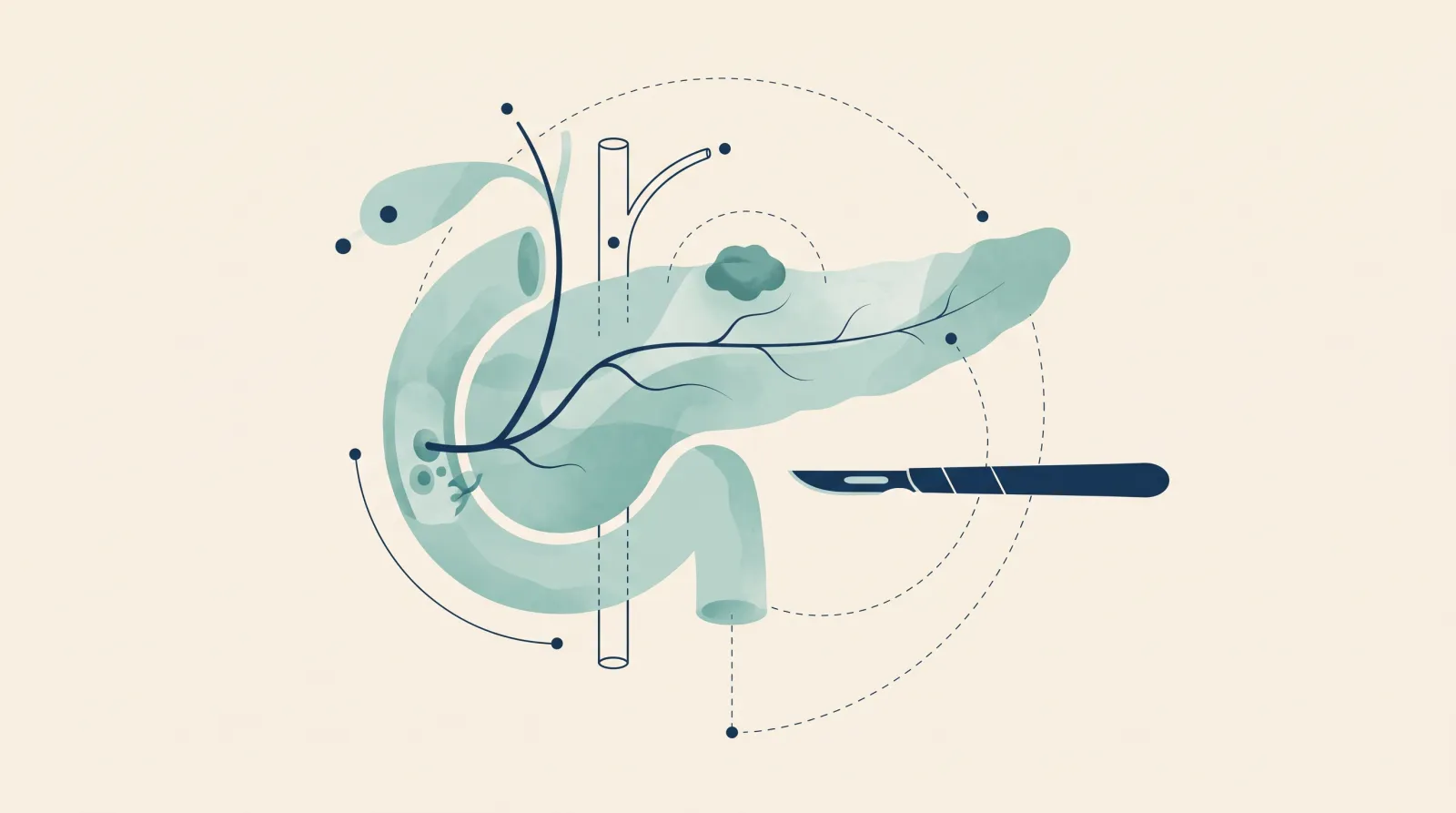
Records. PET-CT · biopsy reports with subtype, CD19 / CD20 / CD30 status and MYC / BCL2 / BCL6 status · treatments so far with dates, including the CAR-T product and infusion date, and the bispecific and its last dose. The physician confirms the list per case.

Setting. Most procedures are outpatient under conscious sedation; general anesthesia or an overnight stay is rarely required. Needle procedures take about an hour with about two hours' recovery.

Follow-up. PET-CT at 6 to 8 weeks and blood tests; the number of treatments is decided with the patient's physician; the patient's own hematologist is kept informed throughout.

Dr. Jason R. Williams
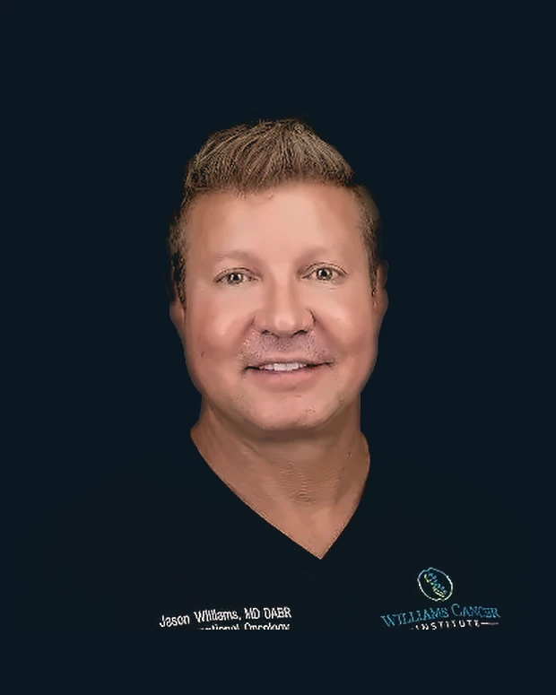
Jason R. Williams, MD, DABRInterventional oncology
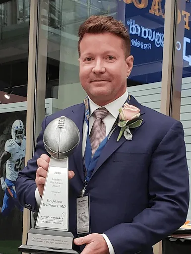
Dr. Williams holding the 2019 Vince Lombardi Cancer Foundation Leaders For A Cure award
Leaders For A Cure2019 · Vince Lombardi Cancer Foundation
The physician who decides every case

Dr. Jason R. Williams

MD, DABR · Director of Interventional Oncology and Immunotherapy

Dr. Jason Williams is a board-certified radiologist who has spent his career putting needles into tumors under imaging. His first patent, in 2003, covered ablation combined with immunotherapy injected into the tumor. A physician since 2000, he opened his first ablation center in 2004 and completed his residency in 2007. He also co-founded Syncromune, whose separate investigational drug SV-102 holds FDA Fast Track status. His tumor board reviews every case.

The team

Meet Our Medical Team

Tap a photo to read their background.
{{ teamSel.r }}

{{ teamSel.n }}

{{ para }}

Where treatment happens

Our Treatment Facilities in Los Cabos

Most of our treatments are performed in Los Cabos, Mexico, in a private hospital setting.

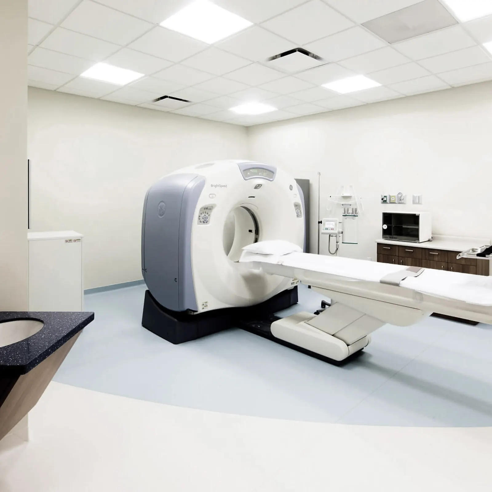
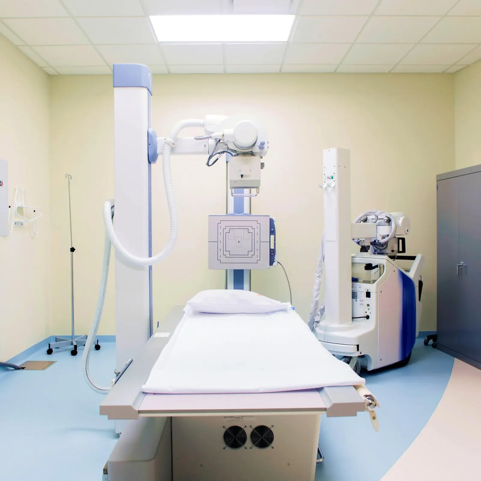
  • Imaging on site, including CT, ultrasound, MRI and digital radiology, for image-guided procedures.
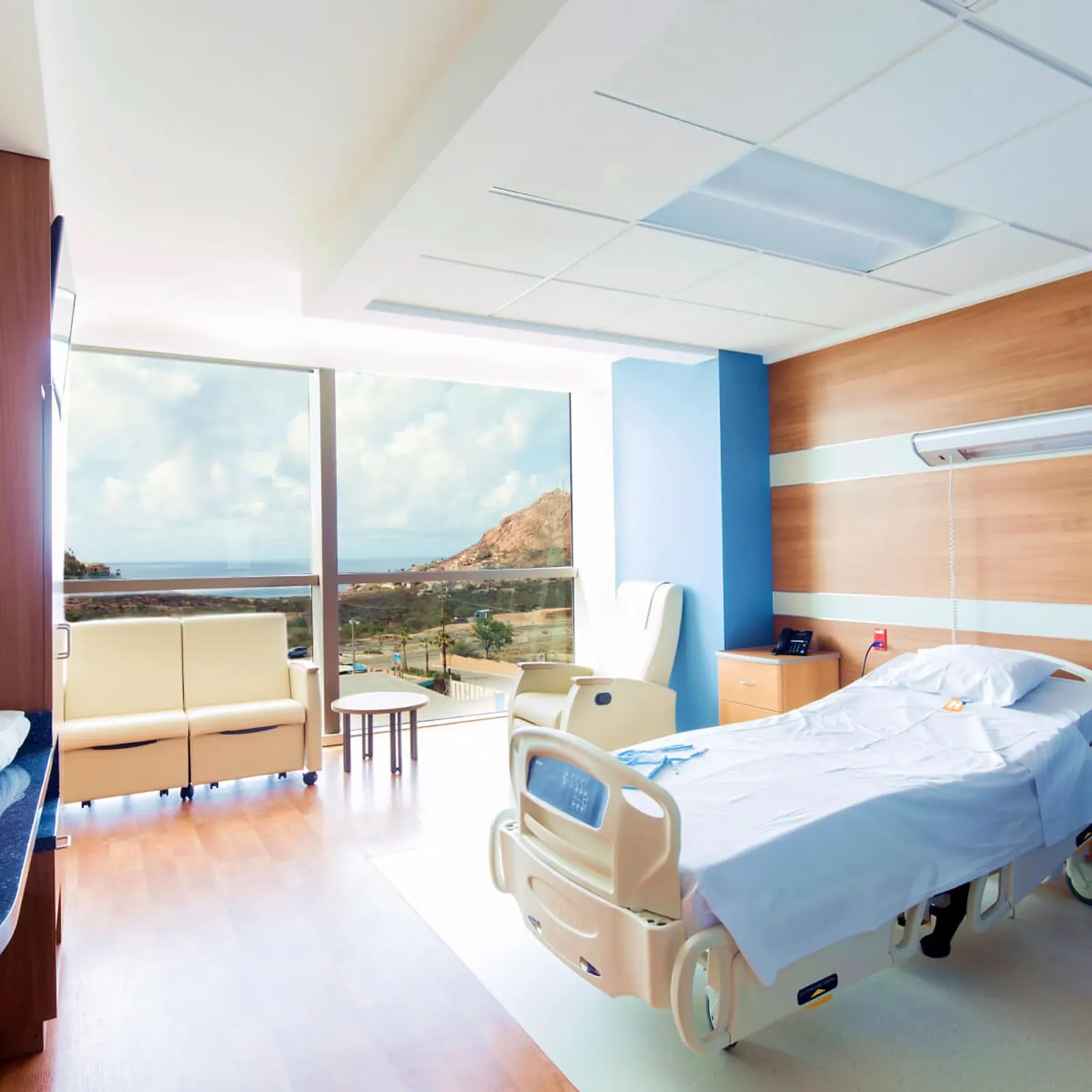
  • Private patient rooms. When an overnight stay is necessary, nursing care is available on the inpatient floor.
  • Located approximately 15 minutes from Los Cabos International Airport (SJD).

Other treatment locations. In some cases our medical team may schedule a procedure in Mexico City. Rebyota is given only at our Beverly Hills, California location.

Aerial view of the Los Cabos marina at sunset
Los Cabos
Waiting lounge overlooking the Sea of Cortez
Patient lounge
Private patient room with ocean view
Private room
CT procedure suite
Procedure suite
Digital radiology room
Radiology
Patient accounts

In their own words

Accounts Williams Cancer Institute publishes, shared with each patient's permission. They span different cancers. Individual results vary and are not guaranteed.

Videos

Lymphoma on video

Selected Lymphoma videos from the Williams Cancer Institute channel. Patient stories are shared with each patient's permission. Individual results vary.
The full Lymphoma playlist on the Williams Cancer Institute YouTube channel.
Next step

Start Your Admissions Process

Complete our admissions form to get started. One of our Patient Consultants will contact you to explain the process and guide you through the next steps, including the medical information and imaging studies required for our medical team to evaluate your case.
info@williamscancerinstitute.com

Scan image not available in this build{{ zoomCap }}
{{ zoomCap }}