Williams Cancer Institute +1 (863) 451-9755
Breast Cancer

Breast cancer, treated through a needle where it sits.

We break the tumor open through a needle, with electrical pulses or freezing, and inject immunotherapy into that spot, so your immune system can learn what to hunt. For breast cancer that is early, or that the drugs no longer touch.

Start Your Admissions Process How It Works

Complete a short, three-step form to begin your admissions process. One of our patient consultants will contact you to explain the process and guide you through the next steps.

What we do
AblationVaxTreat the tumor. Teach the immune system. Treat locally, act globally.Everything we do is built on AblationVax, the approach Dr. Williams developed: the tumor is treated through a needle (ablation), and the treated tumor becomes an in-situ vaccine: a vaccine made inside your body, from your own cancer.

Work from inside the tumor, not from outside

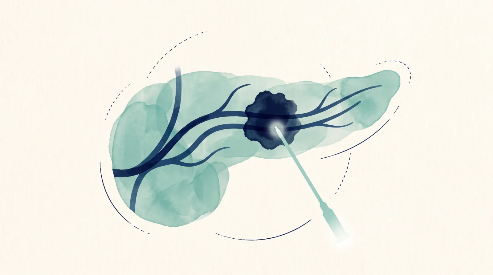
Most breast cancer care reaches the tumor from outside: surgery removes it, drugs travel through your body. We go in directly. Under ultrasound guidance, a WCI physician places a thin needle into the tumor and breaks its cells open with electrical pulses or freezing, releasing the tumor's own antigens: the markers the immune system uses to recognize cancer. In the same session, immune drugs go into that exact spot. It works alongside your current treatment, not in place of it.

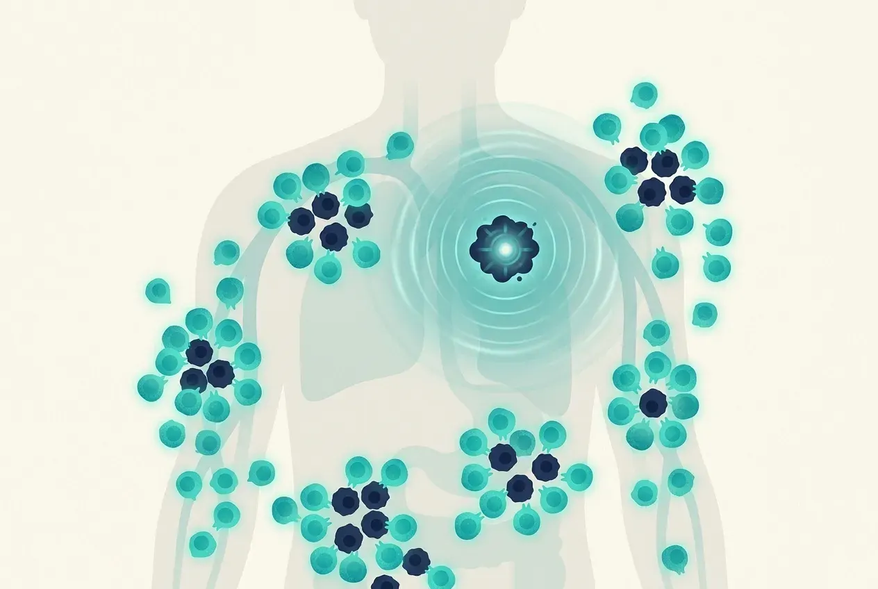
The immune drugs placed in the tumor teach the immune system to recognize this cancer, the way a vaccine does. You teach the immune system in one spot, and it goes after the same cancer in the rest of the body.

1Break it openA needle breaks the tumor open with electrical pulses or freezing, releasing its antigens.
2Treat it in placeImmune drugs go into that exact spot, in the same session.
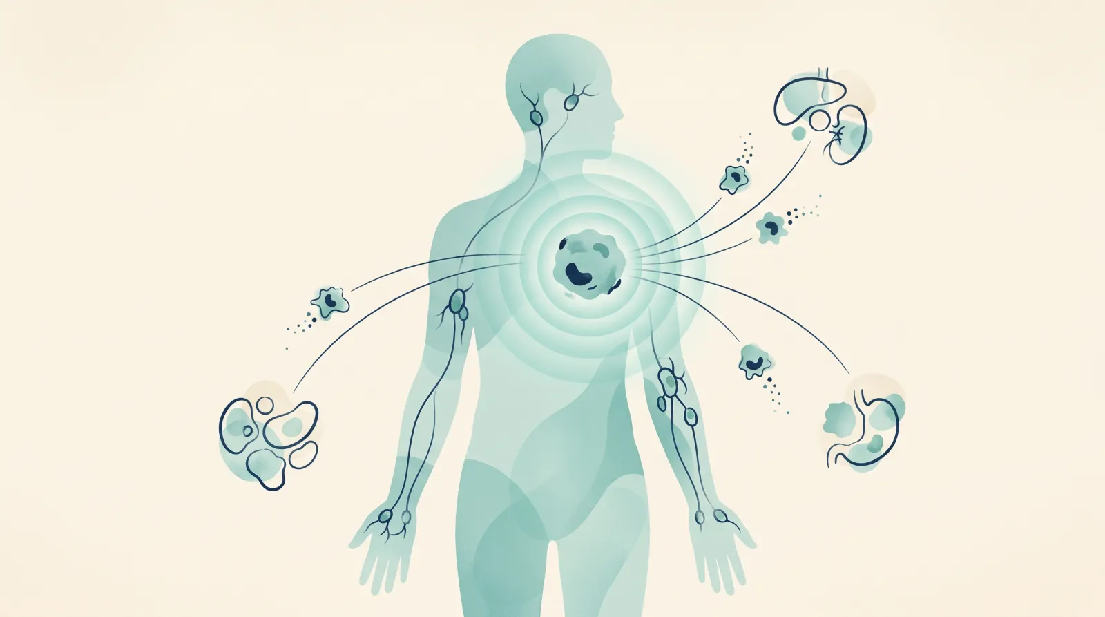
3Teach the whole bodyThe immune system learns the cancer at one tumor and goes after it everywhere else. Doctors call this the abscopal effect. It does not stop there: the cycle below keeps it going.
Video (1:22): Why do electricity and immunotherapy work so well together? Dr. Williams explains AblationVax.
After step 3: how AblationVax keeps working

The intratumoral immune conversion cycle

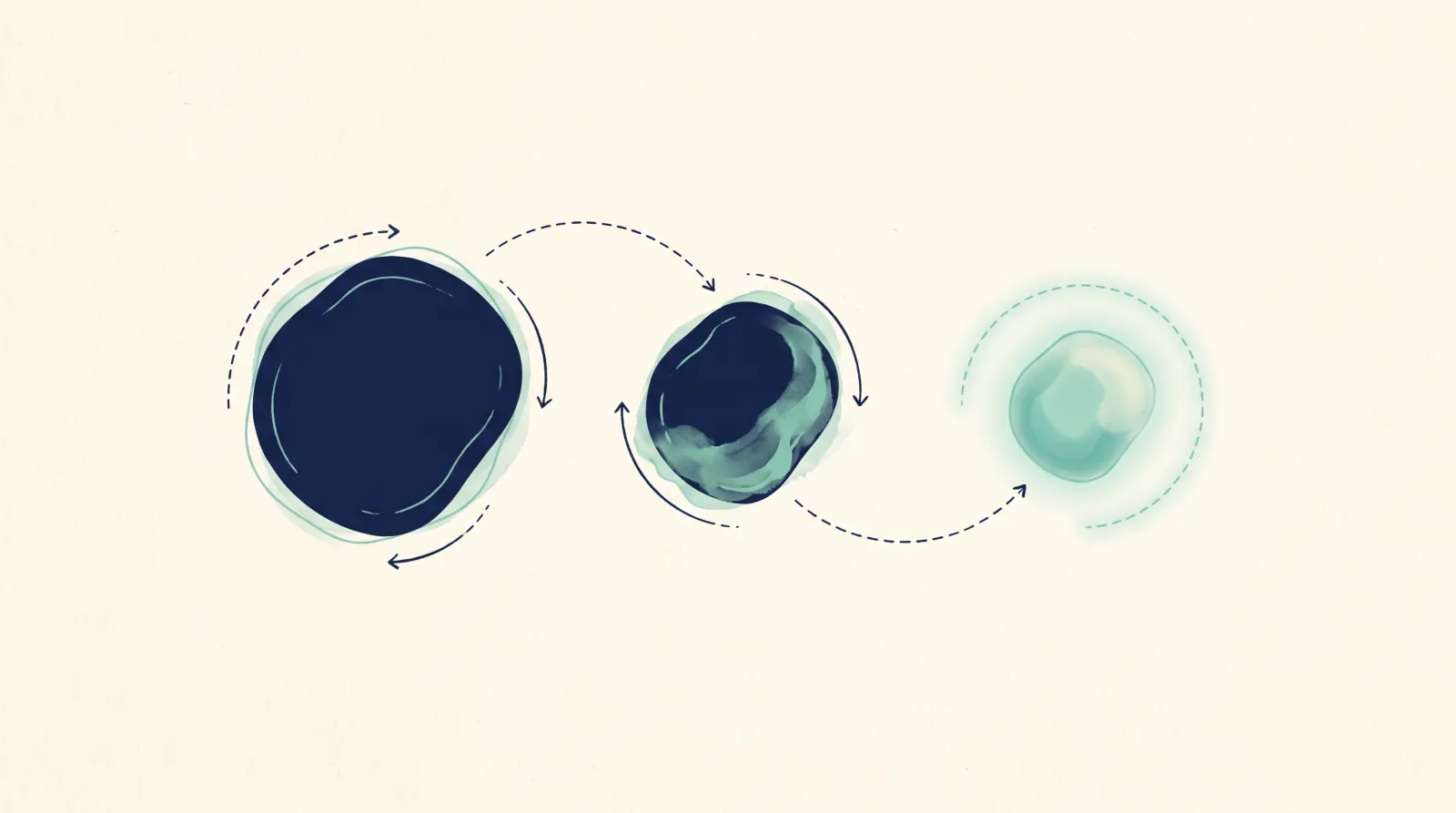
The treated tumor is the source of the vaccine, and it keeps supplying it. It is not one exposure and done. Every round of tumor cells the immune system destroys exposes new antigens, and those restart the cycle, so the response keeps spreading: broader, throughout the body, and with memory. It turns a “cold” tumor, one the immune system ignores, “hot”.

01DisruptAblation breaks the tumor open and exposes its antigens, the markers the immune system uses to recognize the cancer: not a few, all of them.
02Prime and recruitImmune drugs in that same spot call immune cells in and show them the cancer.
03Activate and co-stimulateCo-stimulation is a second go-signal. Those immune cells are switched on against the antigens the ablation just exposed.
04ReleaseThe checkpoint drugs we inject take the brakes off, so the immune system can kill tumor cells.
05DestroyImmune cells destroy tumor cells, and every cell they kill exposes new antigens.
06RepeatThe new antigens restart the cycle: re-priming, amplifying and spreading the response.

Card 06 leads back to card 02. Doctors call the widening response epitope spreading.

Most cancer vaccines: outside-inMade outside the body from a few pre-selected antigens, the same for everyone, then injected. The response is limited by the antigens someone chose.
AblationVax: inside-outMade inside your body, from every antigen in your own tumor. A vaccine made for one person: you. Not a patch: a rebuilding of the immune system from within.

A short procedure, not an operation

It takes about an hour, under ultrasound guidance, and most people go home the same day, about two hours later. Freezing a breast tumor through a needle is established enough that, on its own, it is now authorized in the United States for a defined group of older patients with small, low-risk tumors.

Wakes up an immune system that was ignoring the tumor

Many breast tumors are quiet to the immune system. Even triple-negative tumors often slip past checkpoint drugs given on their own. Breaking the tumor open releases its antigens, so the immune system finally has something to see. The drugs we inject into that spot are chosen to wake it up: some release its brakes, others press the accelerator.

Electricity or freezing, chosen per tumor

Pulsed electric fields are used in most cases; freezing (cryoablation) in some, as in the case below. Either way, immune drugs go into the treated spot in the same session.

Reaching cancer it never touched

Once the immune system has been alerted at one tumor and kept re-primed by the cycle above, it goes after the same cancer elsewhere in the body, and it remembers it. Doctors call this an abscopal effect.

Standard care. Surgery, radiation and systemic therapy, chosen by stage and receptor status. Cryoablation on its own is authorized in the United States for one defined group: in October 2025 the FDA granted marketing authorization to IceCure's ProSense system for early-stage, low-risk breast cancer in women aged 70 or older with ER-positive, PR-positive, HER2-negative tumors of 1.5 cm or smaller, used with adjuvant endocrine therapy. That authorization covers cryoablation alone. In triple-negative disease, intravenous (IV) checkpoint blockade gives systemic drug exposure.

Ablation alone. Local treatment of the tumor; any immune effect of ablation alone is not established in patients.

This approach. Image-guided ablation of the breast tumor, by pulsed electric field (PEF) ablation in most cases and cryoablation in some, combined in the same session with a multi-agent intratumoral immunotherapy injected into the ablation bed. It is used across stages and receptor types, under physician review. In our clinical observations, a systemic immune response occurs beyond the treated tumor.

Rationale. Hormone-receptor-positive tumors are usually immune-quiet; triple-negative tumors often do not respond to checkpoint drugs given alone. WCI's rationale is that ablation exposes the tumor's antigens and that the injected agents drive an immune response to them at the site. This is what we see in our clinical observations.

The procedure, step by step

Guidance. Ultrasound-guided percutaneous access to the breast tumor.

Ablation. PEF delivers electrical pulses through the single needle, without heat; where cryoablation is used, cryoprobes form an ice ball around the tumor. Lethal cell kill occurs in the inner zone, where tissue falls below −40 °C. More than one freeze–thaw cycle is used to maximize cell death and antigen release. Dying cells release damage-associated molecular patterns (ATP, HMGB1, calreticulin) and intact protein antigens.

Intratumoral immunotherapy. A multi-agent combination is injected into the ablation bed, through the same needle after PEF or through a second needle after cryoablation. The combination is chosen from the tumor's biology (ER, PR, HER2 including HER2-low, Ki-67, BRCA, PD-L1, TROP-2) and from prior treatment.

Setting and time. About an hour for the procedure under conscious sedation, and about two hours of recovery. Most procedures are outpatient under conscious sedation; general anesthesia or an overnight stay is rarely required.

Follow-up. Imaging at 6 to 8 weeks (PET/CT, MRI or ultrasound as appropriate). Repeat procedures as indicated; the number of treatments is decided with the patient's physician. The patient's home oncology team is kept informed throughout.

Agents. The injected immunotherapy is a multi-agent combination. The agents and doses are set per patient by the treating physician.

The immune conversion cycle, in clinical terms. Ablation disrupts the tumor and exposes its full antigen repertoire, not a pre-selected subset. The injected immunotherapy then remodels the tumor microenvironment, priming, recruiting and activating immune cells and releasing the brakes on them. Immune-mediated lysis exposes further antigens, which re-prime the response (epitope spreading), producing a broader, polyclonal, systemic response with memory, converting a cold tumor hot. This is WCI’s stated rationale, in Dr. Goodyear’s framing, borne out in our clinical observations.

Evidence, and its limits. In mice with colorectal tumors and no ablation, anti-CTLA-4 injected into a tumor reached about 14 times the day-7 level of IV dosing in that tumor; the untreated tumor received no more than with IV dosing, and half the dose reached the blood within an hour [1]. That is mouse data in a colorectal model; it does not test ablation or show a distant-site effect. In a 2024 mouse model of triple-negative breast cancer, cryoablation of the primary tumor altered the immune microenvironment at distant sites [2]. Image-guided breast cryoablation as a technique dates to a 2004 multi-institutional pilot [3]. There is no randomized trial of cryoablation combined with intratumoral immunotherapy in breast cancer. The case under 'What the records show' is a case record, not a series with a denominator.

Sources[1] Meyblum L, et al. J Immunother Cancer 2023. PMID 37949616.[2] Sardela de Miranda F, et al. 2024. PMID 39703517 (mouse model of triple-negative breast cancer).[3] Sabel MS, et al. 2004. PMID 15123465 (multi-institutional pilot, image-guided breast cryoablation).

What the records show

Four breast cases, with the scans

Four patients' courses from our records, with the scans exactly as recorded. Not every patient will have the same result.

Case1
Partial response

Ultrasound-Guided Cryoablation with Intratumoral Immunotherapy for Left Breast Carcinoma, with Subsequent Nodal Recurrence and Re-Treatment

Left breast cancer with subsequent axillary nodal recurrence.

Partial response, after re-treatment of a node recurrence

In plain words

The tumor shrank. It came back in a node, and was treated again.

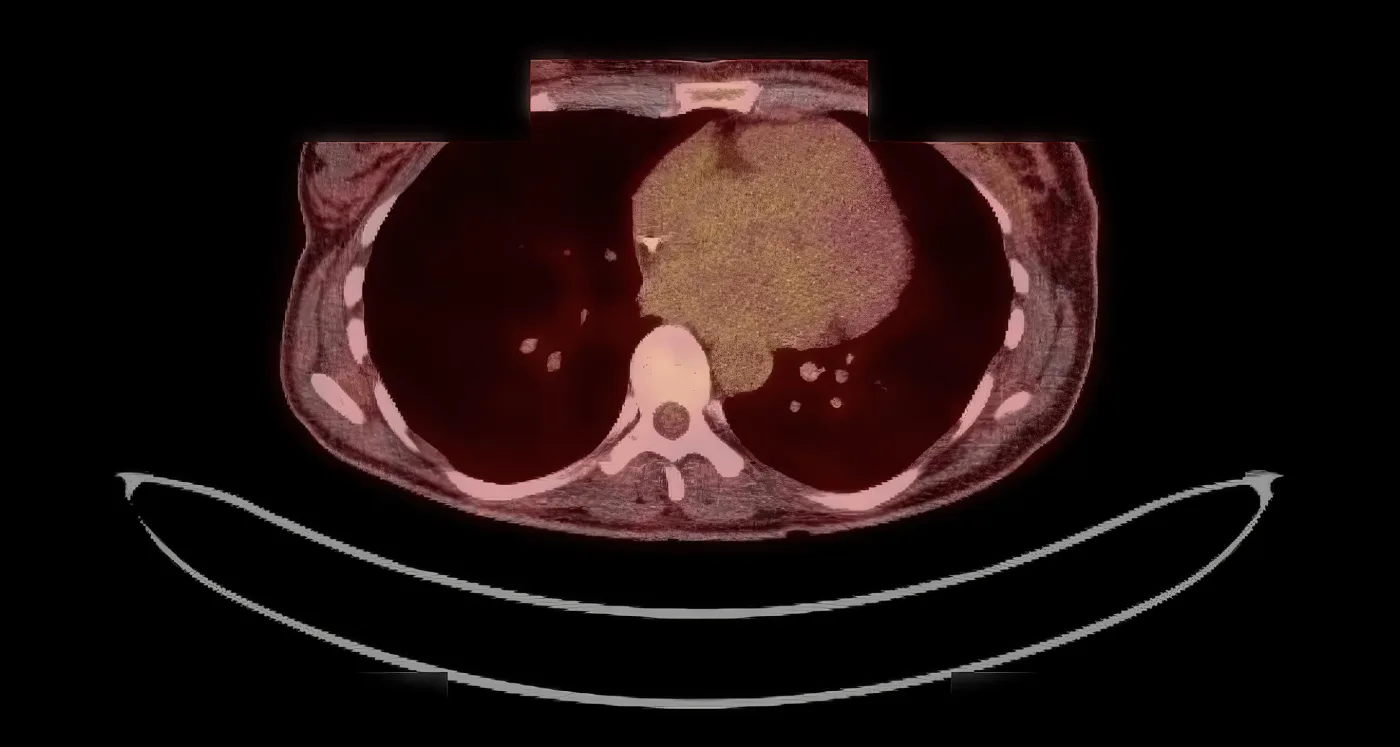
A woman newly diagnosed with left breast cancer, a 3.6 cm tumor. A needle froze the tumor and immunotherapy was injected into it. At month 11 new active lymph nodes appeared under the arm, with a new small spot in the breast, and a second session treated the node. By month 16 the breast tumor was smaller (3.6 × 2.5 cm to 2.4 × 1.2 cm), its activity on the scan was down by about two-thirds, and there was no sign of active spread.

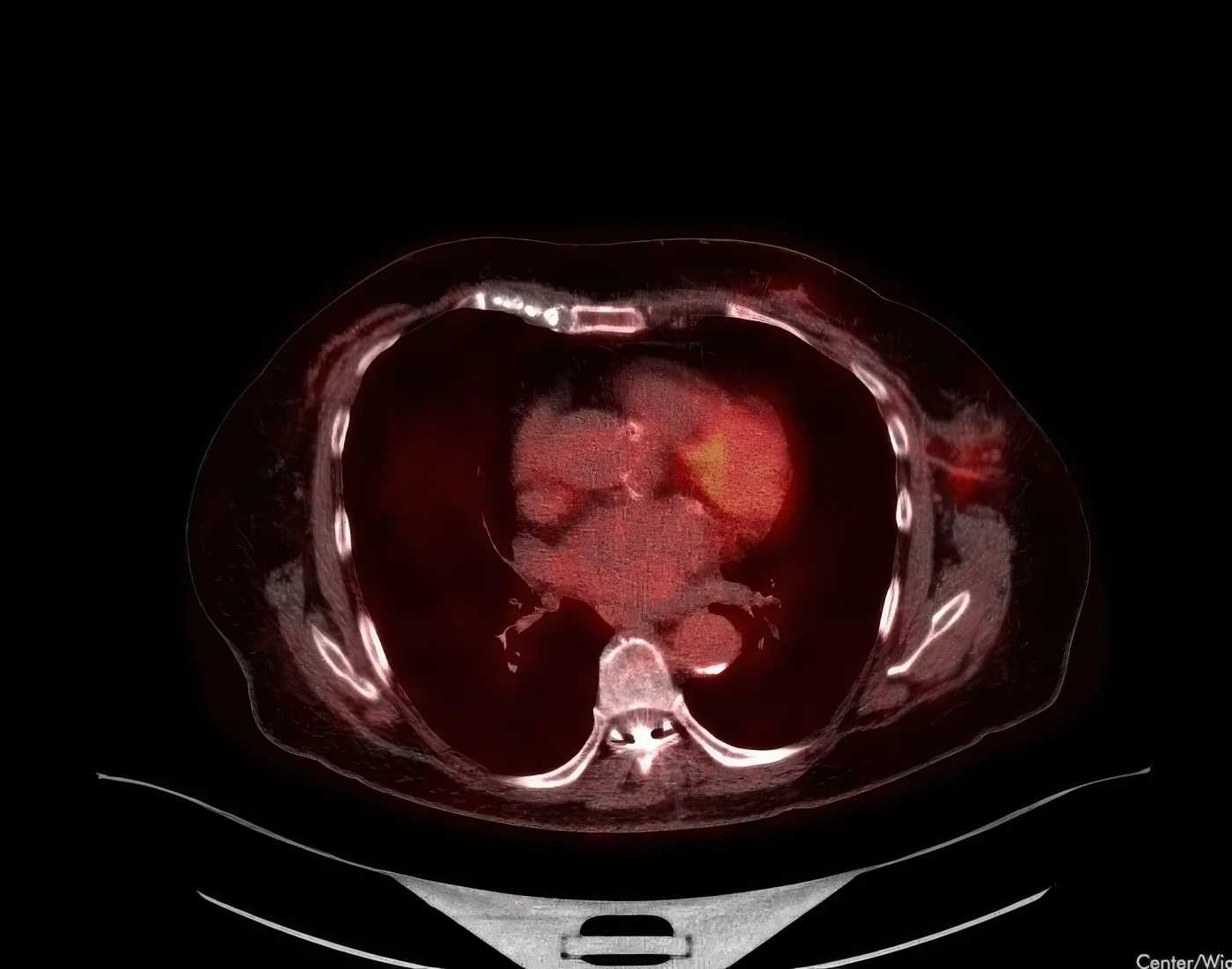
PET/CT, Month 16 · follow-up
December 2023 · Month 16 · PET/CTActivity down 69%
Case study

It came back in the lymph nodes. She was treated again.

The patient

A woman newly diagnosed with left breast cancer. Staging showed a 3.6 cm active tumor reaching from the skin to the chest-wall muscle, with no sign of spread.

What was done

She had ultrasound-guided cryoablation of the breast tumor, two freeze-thaw cycles, with immunotherapy injected into the tumor bed. She attempted a single chemotherapy session and did not tolerate it. At month 11 a restaging scan found the disease had progressed: new active lymph nodes under the left arm, and a new small active spot in the breast. A second session followed, aimed at the node under the arm.

What the record shows

At month 16 the breast tumor had shrunk from 3.6 × 2.5 cm to 2.4 × 1.2 cm, its activity was down from 8.3 to 2.6, and there was no sign of active spread. New small spots in the right lower lung were read as more likely infection or inflammation than cancer. This is a partial response, reached after documented progression and re-treatment.

Following re-treatment of the axillary node, PET/CT in Month 16 showed marked reduction in left breast tumour size and metabolic activity with no evidence of metastatic disease. Note that this course included documented interval progression in Month 11 between the two treatment sessions.

Timeline

Course, measured from the first treatment

Activity · FDG PET
SUV 8.3→2.6
August 2022 → December 2023 · Day −20 → Month 16 · 69% lower
Size · PET/CT
3.4 × 2.3 cm→2.4 × 1.2 cm
December 2023 · Prior → Month 16 · left breast tumor
Before Treatment2 sessions Follow-up
August 2022Day −20Baseline PET
August 2022Day 0Session 1
August 2023Month 11Node · Session 2
December 2023Month 16PET · activity −69%
Treatment session Key scan Baseline imaging Time to scale

{{ ph.title }}

{{ ph.span }}
  1. {{ ev.t }} Milestone
    {{ ev.sub }}
    {{ ev.metric }}MTM/mL

    {{ ev.v }}

{{ ph.scansLabel }}
Total delivered {{ ph.note }}
Case2
Not characterised

Cryoablation with Intratumoral Immunotherapy for Invasive Lobular Carcinoma of the Left Breast with Axillary Metastasis

Invasive lobular carcinoma, Grade 2, left breast with axillary metastasis

In plain words

Lobular breast cancer with lymph-node spread: a substantial early fall in activity, with the long-term outcome not yet established.

This patient had grade 2 invasive lobular carcinoma of the left breast, hormone-receptor positive and HER2 positive, that had already spread to a lymph node under the arm. Our team treated the breast tumor in a single session of cryoablation, with immunotherapy injected into the tumor bed.

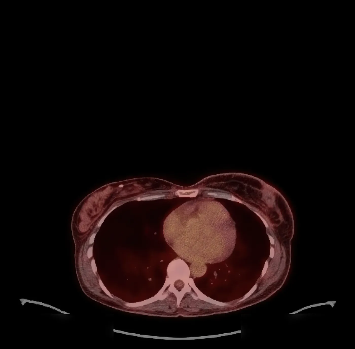
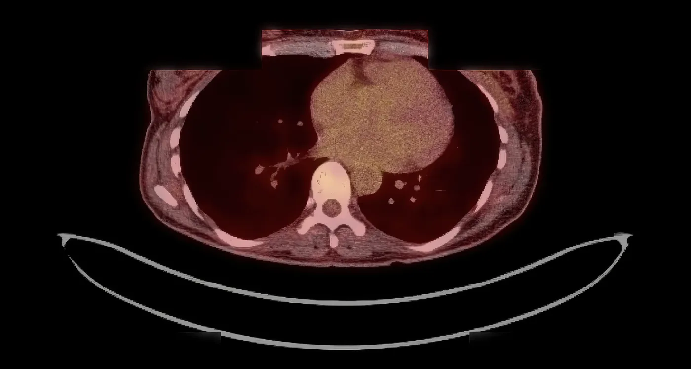
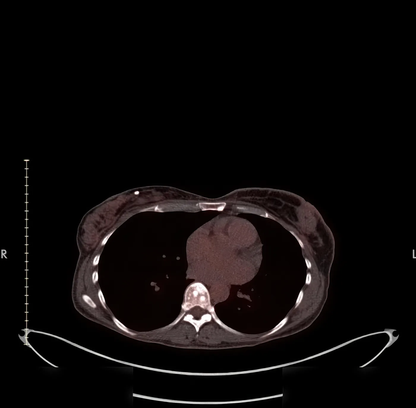
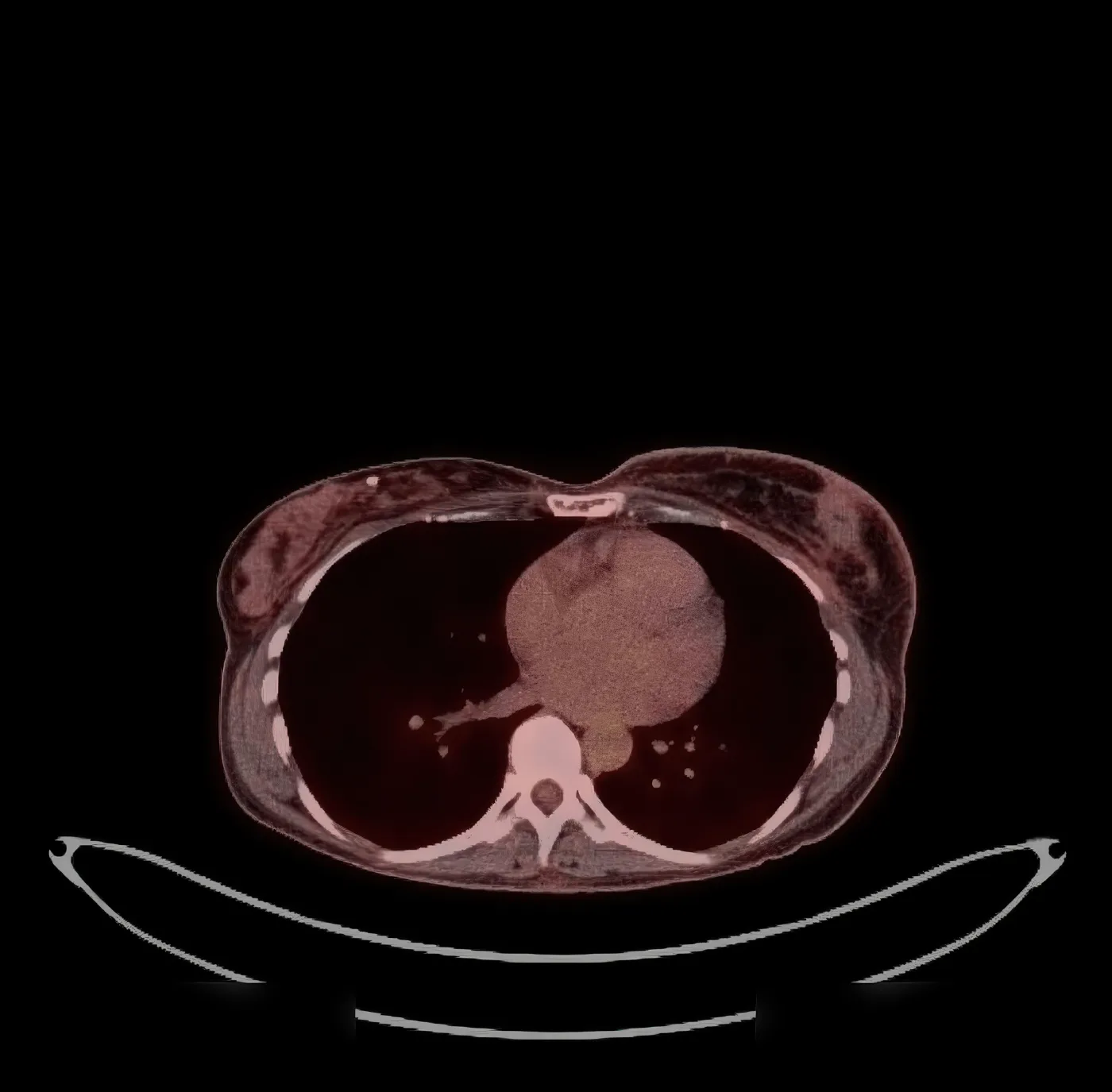
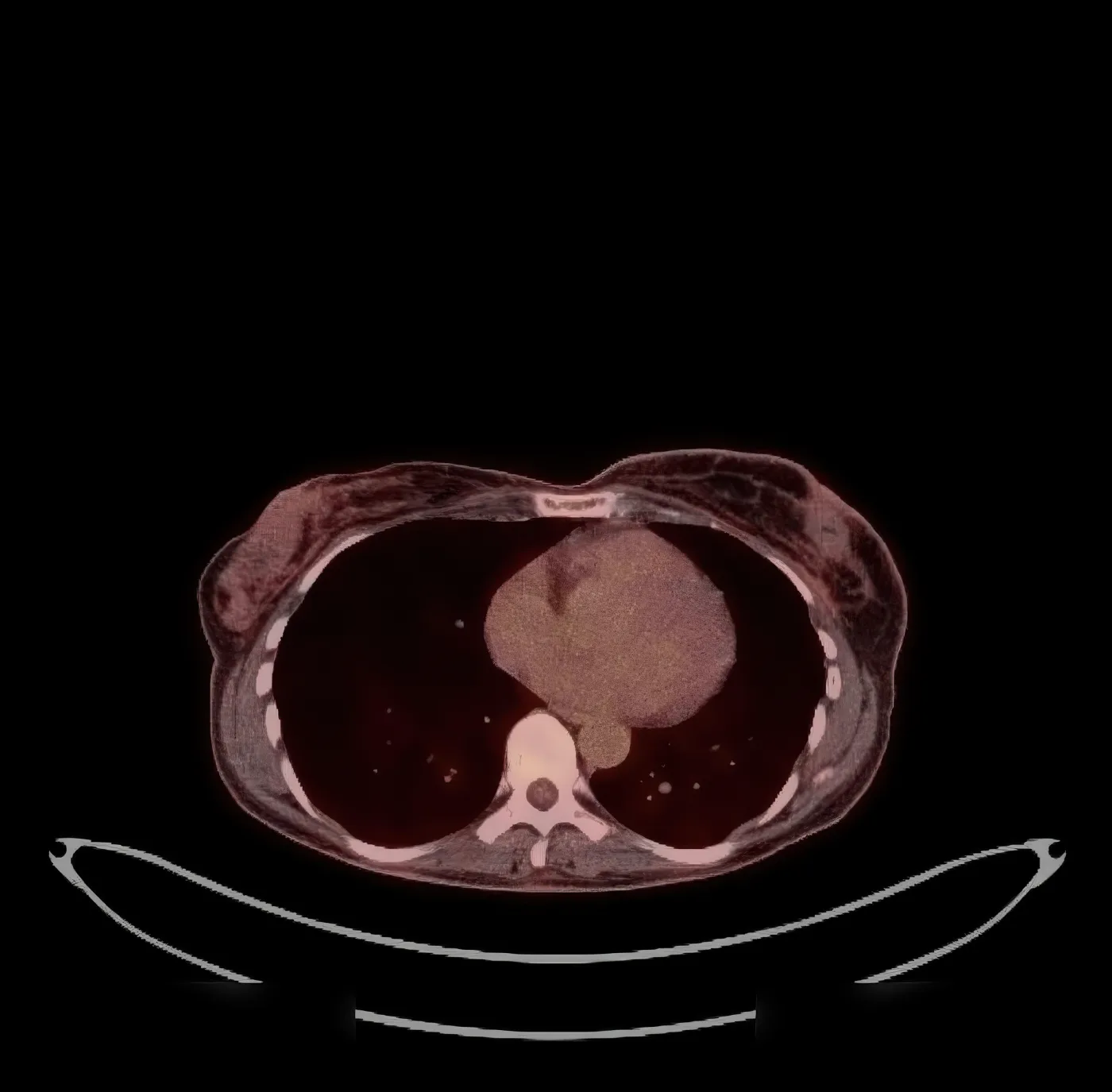
PET/CT · Long-term follow-up · Year 2
December 2024 · PET/CT · Long-term follow-up · Year 2
Case study

A strong early drop. Then scans with no written report.

The patient

A woman with grade 2 invasive lobular carcinoma of the left breast that had already spread to a lymph node under the arm. The tumor was hormone-receptor positive and HER2 positive, measuring 3.5 to 3.7 cm on ultrasound.

What was done

She had a single session of ultrasound-guided cryoablation of the breast tumor, with immunotherapy injected into the tumor bed.

What the record shows

At five and a half months, FDG metabolic activity in the left breast had decreased substantially. The radiologist favoured post-treatment inflammation but could not rule out a small amount of remaining disease. Four later PET/CT images were obtained, out to about two and a half years, and appear in the record without a written radiology report, so no outcome is stated for that period.

One patient's record, assessed retrospectively, with no comparison group. It cannot show this treatment works better than any alternative, and some patients with the same diagnosis do not benefit.

Timeline

Course, measured from the first treatment

March 2022 · Diagnosis (Month −3)Left breast invasive lobular carcinoma, Grade 2. Tumour size at least 1.9 cm. Margins extensively involved by invasive carcinoma. No DCIS identified; no lymphovascular invasion. Pathologic stage at least pT1c, pNx.
Biomarkers (immunohistochemistry)ER positive (22%). PR positive (69%). HER2 by IHC positive (3+). Ki-67 22%. E-cadherin negative, consistent with lobular carcinoma.

April 2022 · Axillary lymph node (Month −2)Left axillary node biopsy showed metastatic carcinoma compatible with breast primary (CKAE1/A3 positive).
Contralateral breastRight breast biopsy negative for carcinoma; benign keloidal fibrosis, possible PASH or lymphocytic mastopathy.
Pre-treatment ultrasoundMeasurable mass approximately 3.5-3.7 cm.
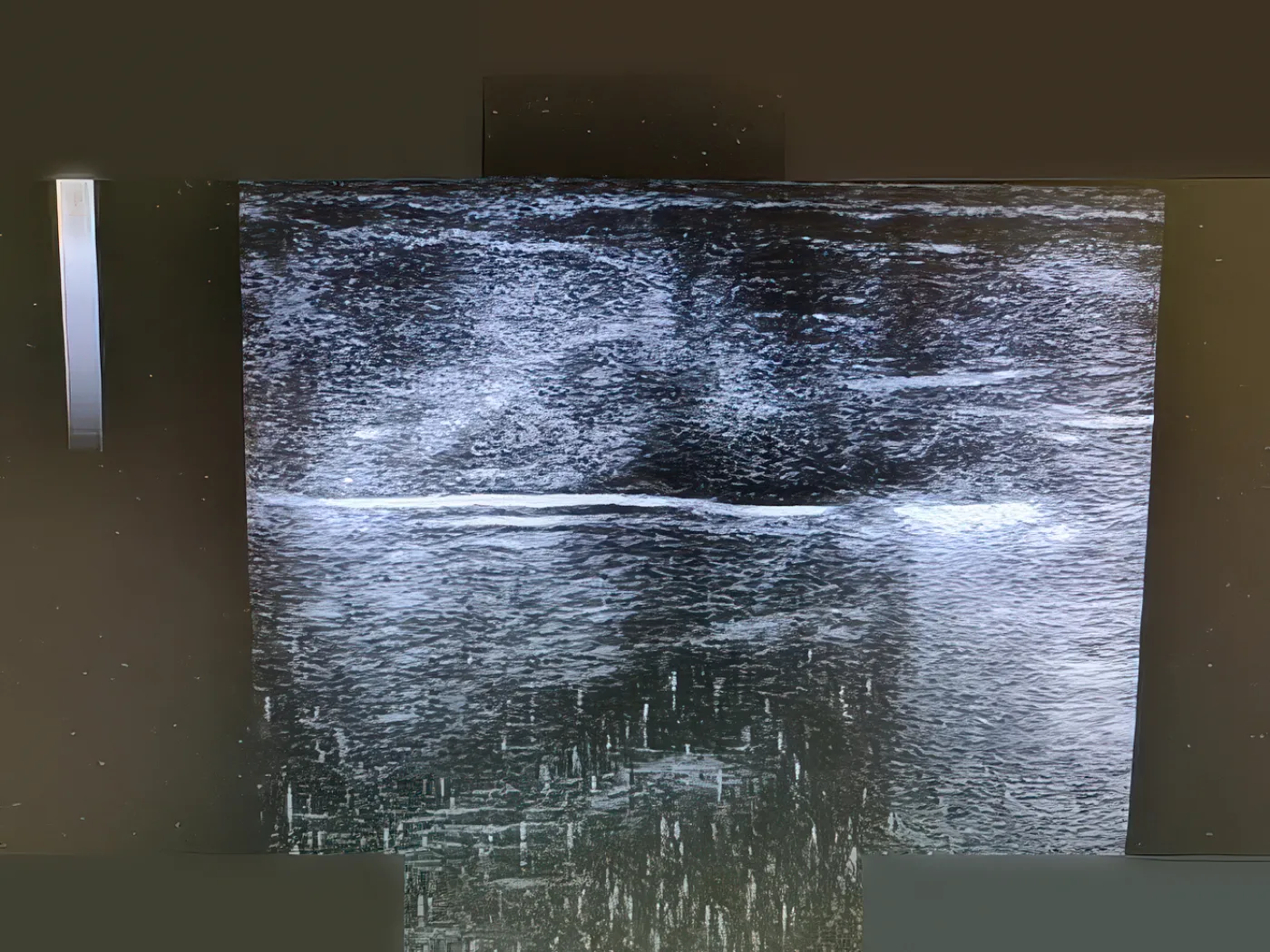
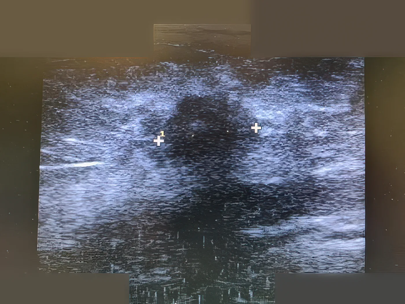
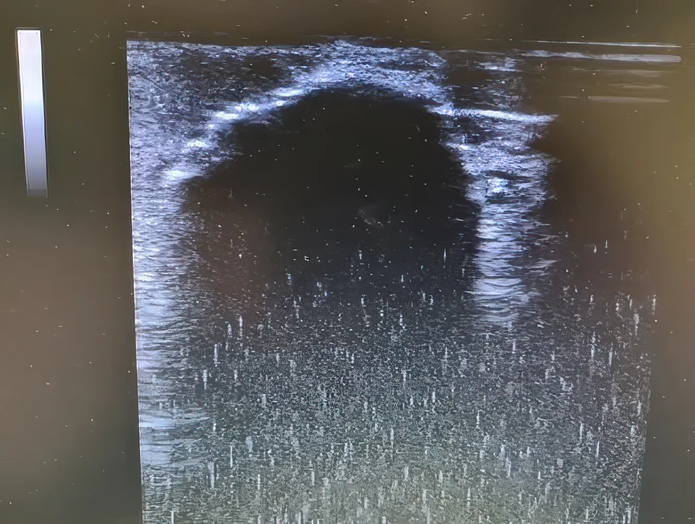
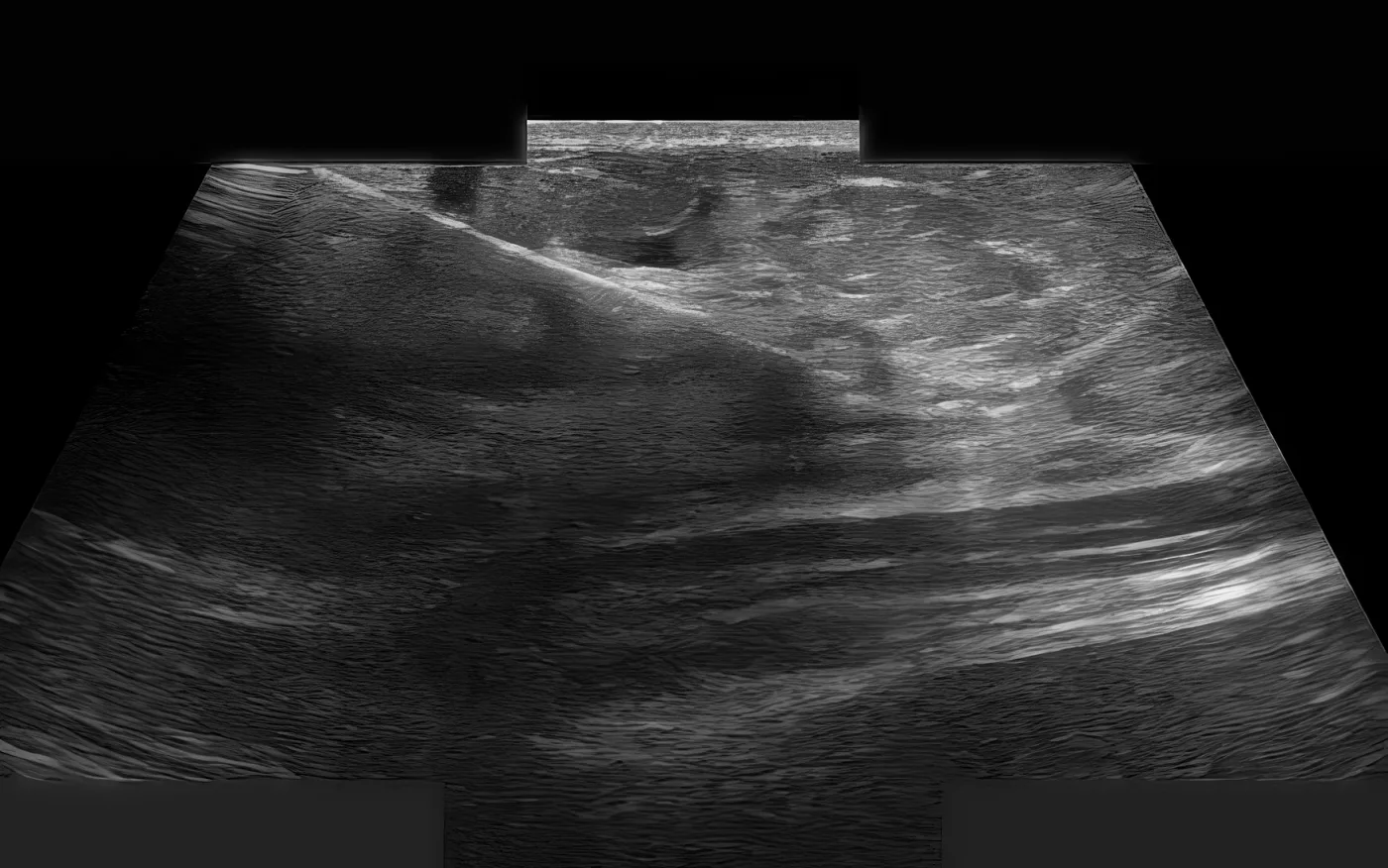
June 2022 · Session #1 · Day 0Ultrasound-guided cryoablation of the breast lesion with intratumoral immunotherapy injection. Intra-procedural ultrasound images document probe placement and ice-ball formation.
Total deliveredOne cryoablation session with intratumoral immunotherapy.
December 2022 · Month 6 · F-18 FDG PET/CT (5.5 months post-treatment)Prominent though significantly less conspicuous metabolic activity localising to left breast skin thickening and along the periphery of less extensive nodular densities in the left breast, current SUV max 2.0. Similar-appearing though non-FDG-avid nodular densities in the right breast. IMPRESSION: significantly less conspicuous activity localising to parenchymal nodules in the left breast. Although the finding favours post-therapeutic inflammatory change, low-volume residual disease could not be definitively excluded.
June 2023 · Month 12 · PET/CTLong-term follow-up imaging obtained. The record presents the images without a written radiology impression.
September 2023 · Month 15 · PET/CTLong-term follow-up imaging obtained. The record presents the images without a written radiology impression.
February 2024 · Month 20 · PET/CTLong-term follow-up imaging obtained. The record presents the images without a written radiology impression.
December 2024 · Year 2.5 · PET/CTMost recent imaging in the record. Presented without a written radiology impression.
Imaging from the record
Ultrasound · Baseline, before treatment
Ultrasound guidance · Session 1 · Day 0
June 2022 · Ultrasound guidance · Session 1 · Day 0
Ultrasound guidance · Session 1 · Day 0
June 2022 · Ultrasound guidance · Session 1 · Day 0
Ultrasound guidance · Session 1 · Day 0
June 2022 · Ultrasound guidance · Session 1 · Day 0
PET/CT · Follow-up · Month 5.5
December 2022 · PET/CT · Follow-up · Month 5.5
PET/CT · Follow-up · Month 5.5
December 2022 · PET/CT · Follow-up · Month 5.5
PET/CT · Long-term follow-up · Year 1
June 2023 · PET/CT · Long-term follow-up · Year 1
PET/CT · Long-term follow-up · Year 1
September 2023 · PET/CT · Long-term follow-up · Year 1
PET/CT · Long-term follow-up · Year 2
February 2024 · PET/CT · Long-term follow-up · Year 2
PET/CT · Long-term follow-up · Year 2
December 2024 · PET/CT · Long-term follow-up · Year 2
Case3
Stable disease

Ultrasound-Guided Cryoablation with Intratumoral Immunotherapy for Invasive Lobular Carcinoma of the Left Breast

Invasive lobular carcinoma of the central left breast

In plain words

Lobular breast cancer held stable, with activity falling below background and no new spread at 13 months.

This patient had invasive lobular carcinoma in the central left breast: a 2.3 cm mass with only mild activity and no spread at staging. Our team treated the tumor in a single session of cryoablation, with immunotherapy injected into the tumor bed.

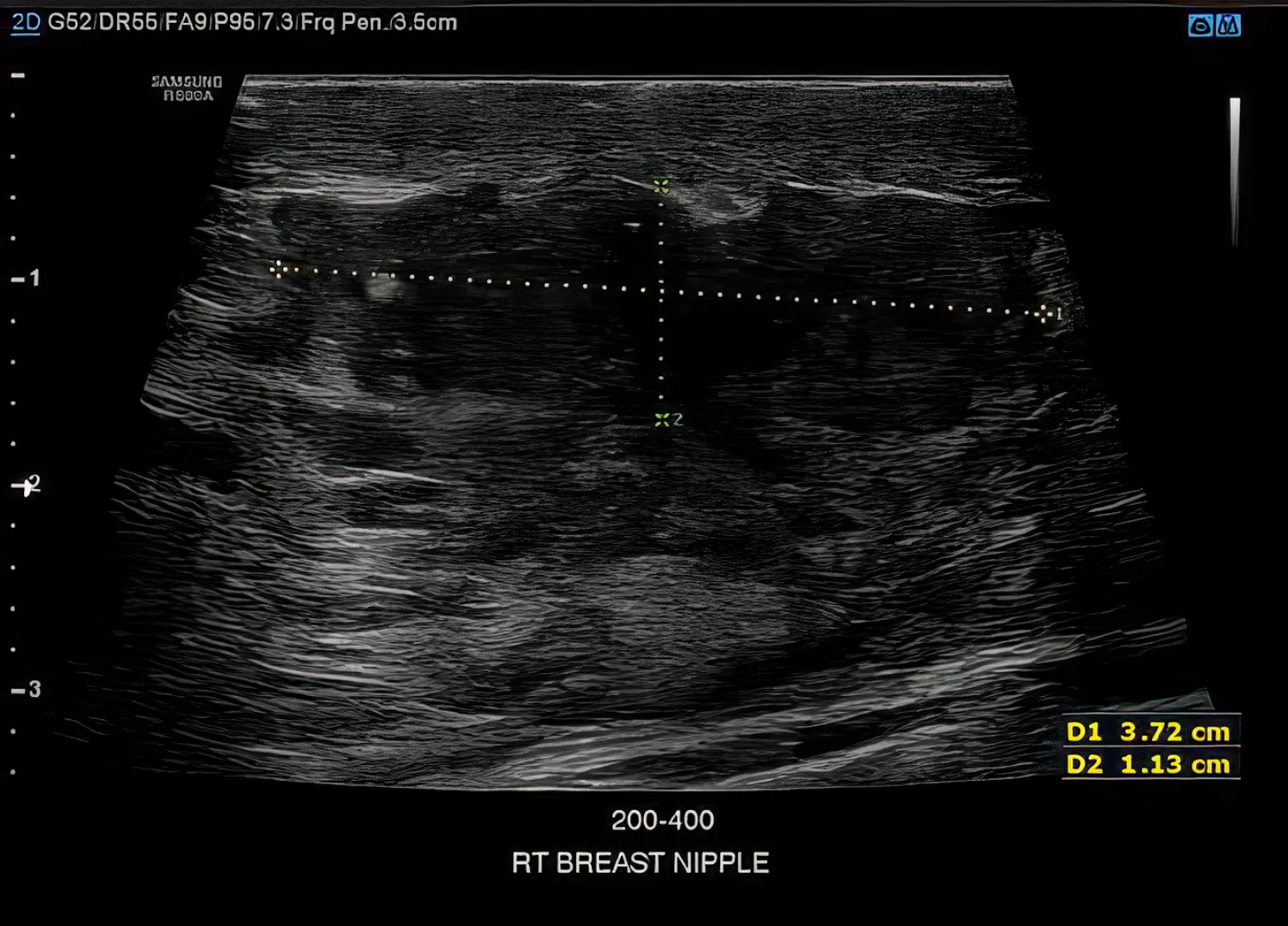
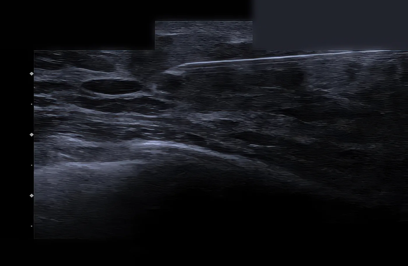
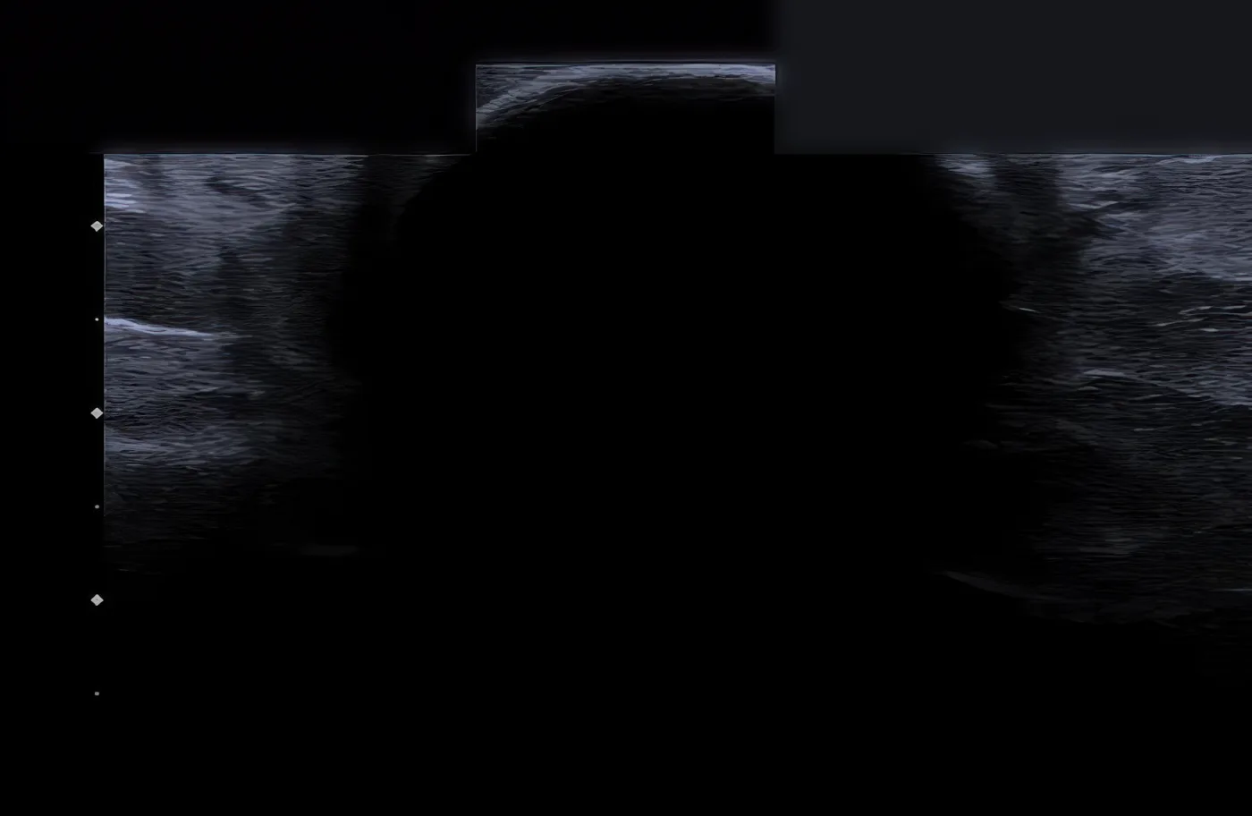
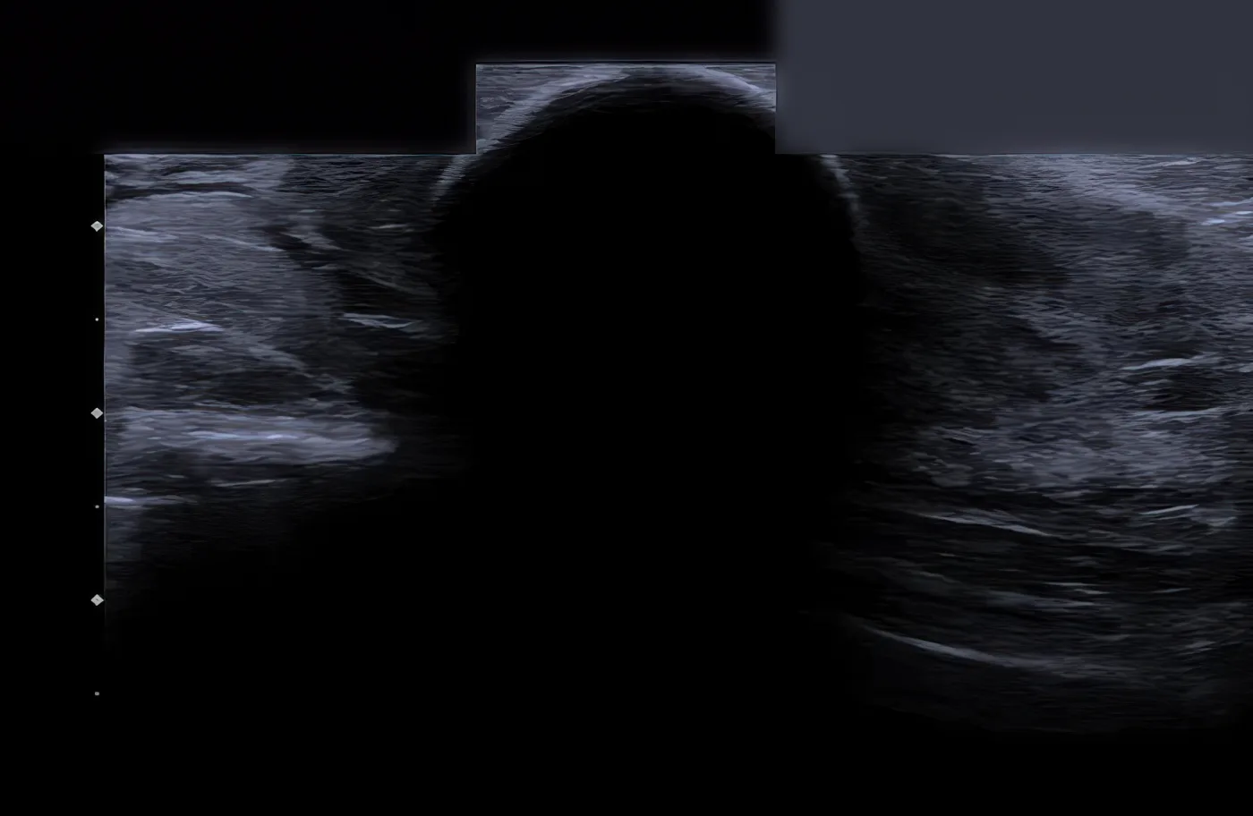
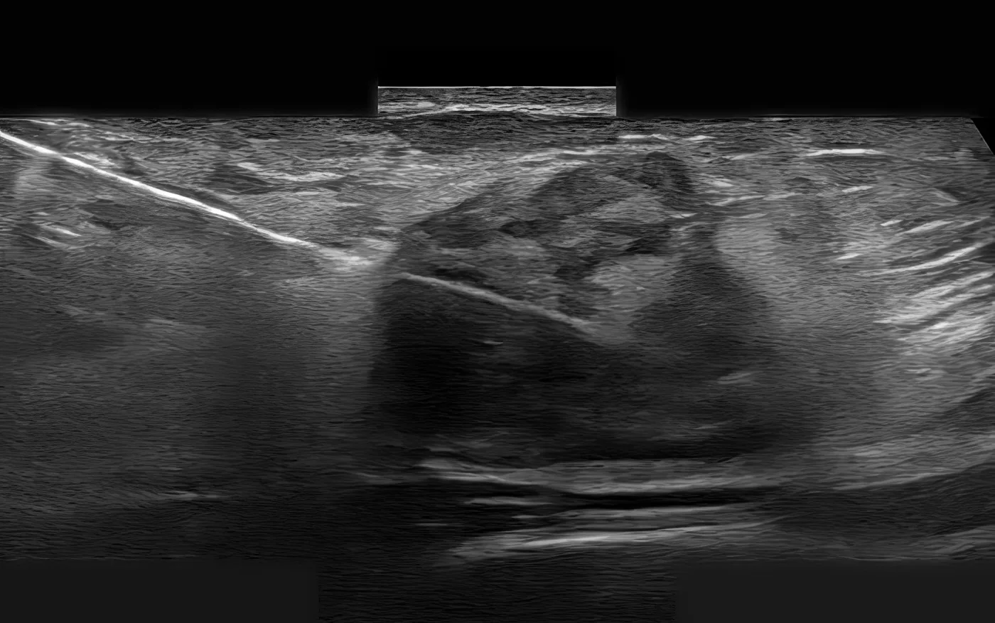
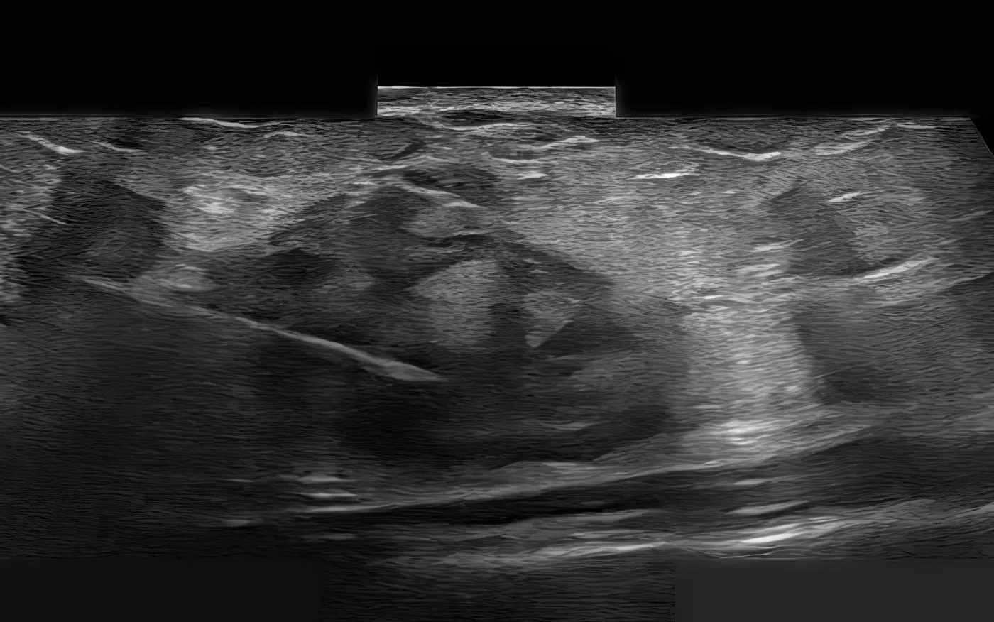
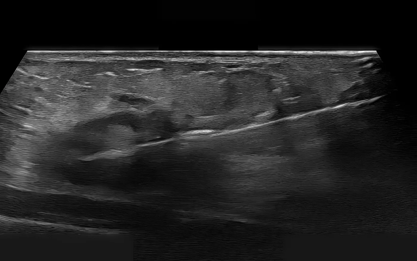
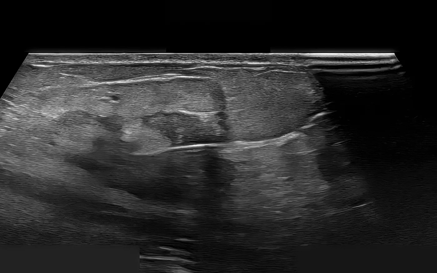
Ultrasound guidance · Session 1 · Day 0
May 2022 · Ultrasound guidance · Session 1 · Day 0
Case study

Not gone. Quieter, and not spreading.

The patient

A woman with invasive lobular carcinoma in the central left breast, 1.4 cm on biopsy. Staging showed a 2.3 cm mass with only mild activity and no spread.

What was done

She had a single session of ultrasound-guided cryoablation of the breast tumor, with immunotherapy injected into the tumor bed.

What the record shows

At about eight and a half months the breast activity was essentially unchanged, with no new disease. By month 13 the mass had fallen below the background activity of the blood, with no new spread. Low-level metabolically active spots in one rib and one vertebra remained stable, with resolution of significant metabolic activity. This is stable disease with decreasing FDG activity, not a complete response.

One patient's record, assessed retrospectively, with no comparison group. It cannot show this treatment works better than any alternative, and some patients with the same diagnosis do not benefit.

Timeline

Course, measured from the first treatment

DiagnosisMalignant neoplasm of the central portion of the left breast.
February 2022 · Pathology (core biopsy Month −3)Infiltrating lobular carcinoma, intermediate histologic grade, 1.4 cm on core. IHC: GATA-3 positive, E-cadherin negative, consistent with lobular features.
February 2022 · Baseline PET/CT (Month −3)2.3 x 2.2 cm left breast mass with mild FDG uptake, SUV 2.87. Small left axillary nodes without abnormal FDG avidity. No distant metastasis.
ImpressionLeft breast invasive lobular carcinoma, clinical stage cT2N0M0 by imaging. No evidence of metastatic disease on staging PET/CT.
May 2022 · Session #1 · Day 0Ultrasound-guided breast lesion cryoablation with intratumoral immunotherapy.
Total deliveredOne cryoablation session with intratumoral immunotherapy.
January 2023 · Month 9 · PET/CT restagingLeft breast: persistent low-level FDG uptake, SUV approximately 2.8-3.0, stable with slight interval change. Left axillary node: low-level uptake, likely post-procedural. Bone: stable low-level uptake at left 6th rib and T7, no new lesions. IMPRESSION: no new FDG-avid metastatic disease.
June 2023 · Month 13 · PET/CTLeft breast mass: SUV now below background blood pool, improved from prior image. No evidence of liver metastasis. Osseous lesions: stable distribution, now without significant SUV activity. IMPRESSION: stable disease, no new metastases.
Imaging from the record
Ultrasound guidance · Session 1 · Day 0
May 2022 · Ultrasound guidance · Session 1 · Day 0
Ultrasound guidance · Session 1 · Day 0
May 2022 · Ultrasound guidance · Session 1 · Day 0
Ultrasound guidance · Session 1 · Day 0
May 2022 · Ultrasound guidance · Session 1 · Day 0
Case4
Not characterised

Pulsed Electric Field Ablation with Intratumoral Immunotherapy for Invasive Ductal Carcinoma of the Right Breast, with Negative Post-Treatment Biopsy

Right breast invasive ductal carcinoma, Grade III, ER+/PR+/HER2-

In plain words

Aggressive breast cancer in which MRI and biopsy disagree after treatment; the response is not yet determined.

This patient had grade III invasive ductal carcinoma of the right breast, hormone-receptor positive and HER2 negative, which grew to at least 6.3 cm with new satellite lesions before treatment. Over four months, our team performed three sessions of ultrasound-guided pulsed electric field ablation, injecting immunotherapy into the tumor each time.

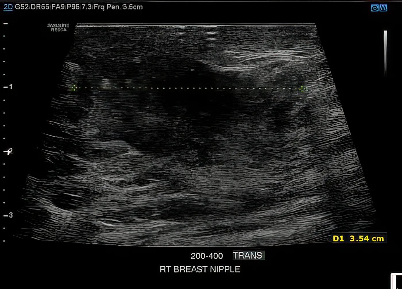
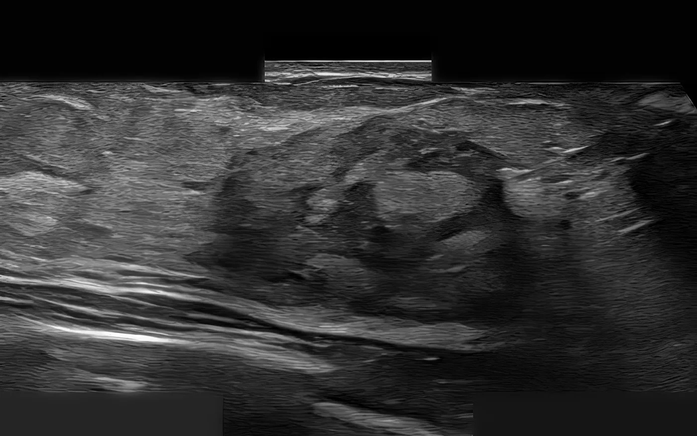
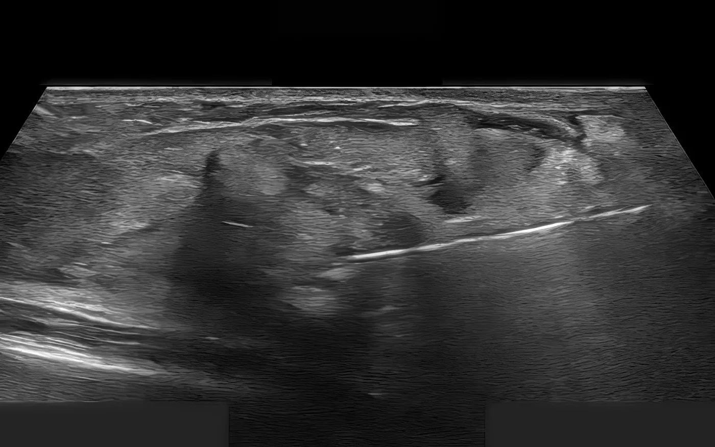
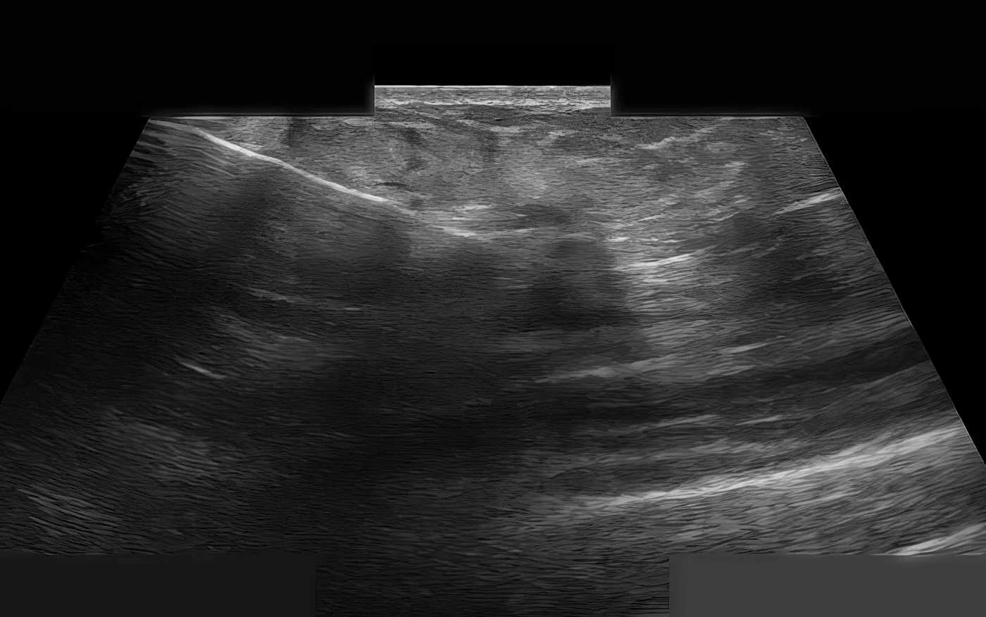
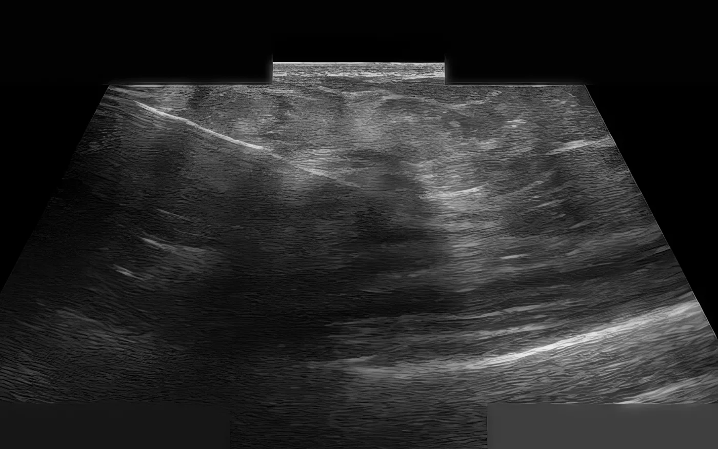
Ultrasound guidance · Session 3 · Month 4
May 2025 · Ultrasound guidance · Session 3 · Month 4
Case study

The MRI and the biopsy disagree.

The patient

A woman with grade III invasive ductal carcinoma of the right breast, hormone-receptor positive and HER2 negative. In the months before treatment the mass grew to at least 4.5 × 3.5 × 6.3 cm, with new satellite lesions around it.

What was done

Over four months she had three sessions of ultrasound-guided pulsed electric field ablation, five cycles in total, with immunotherapy injected into the tumor each time.

What the record shows

At month 10, an MRI was read as persistent viable tumor, slightly larger than before, and surgery was recommended. At month 12 a core needle biopsy found atypical cells and chronic inflammation, and no cancer. The record does not reconcile the two. Sampling error, post-ablation response change and a true response all fit, so no response category is asserted.

One patient's record, assessed retrospectively, with no comparison group. It cannot show this treatment works better than any alternative, and some patients with the same diagnosis do not benefit.

Timeline

Course, measured from the first treatment

January 2022 · Presentation (Year −3.0)Right breast lump. Mammogram showed a 1.2 cm irregular mass, BIRADS 5.
February 2022 · Pathology (Year −2.9)Right breast invasive ductal carcinoma, Grade III, ER positive, PR positive, HER2 negative.
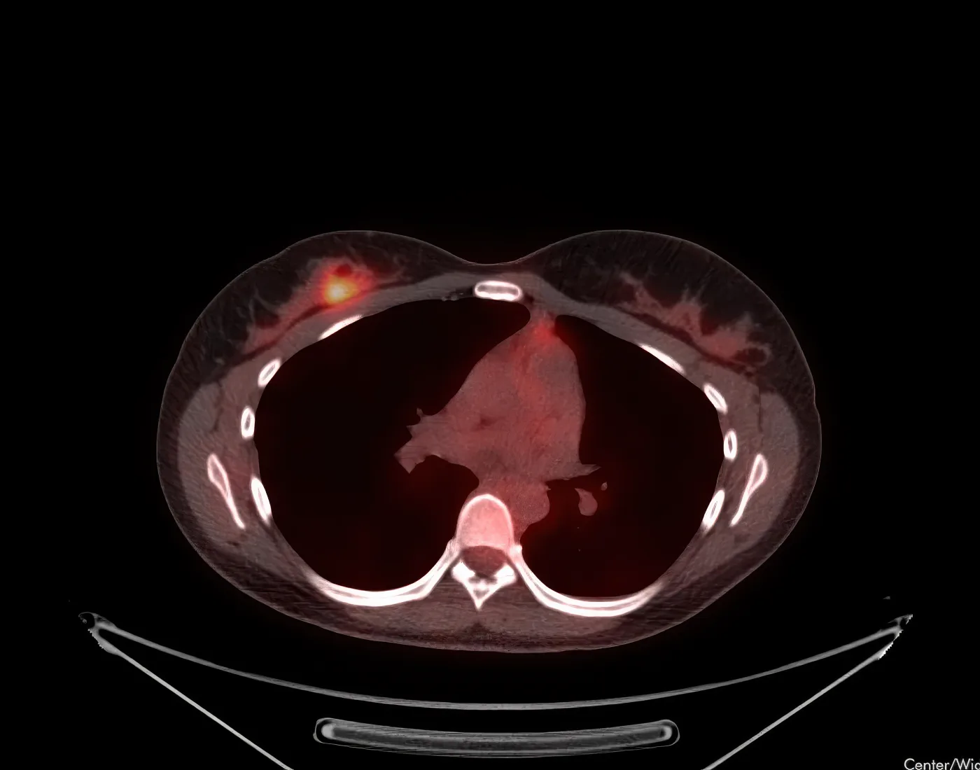
April 2024 · Restaging PET/CT (Month −10)Multifocal FDG avidity in the upper inner quadrant of the right breast, SUV up to 4.9. No regional nodal or distant metastasis identified.
Prior therapy (per the clinic)Adriamycin and Taxol (2022) for 8 months, with a partial response. On Verzenio at the first WCI appointment.
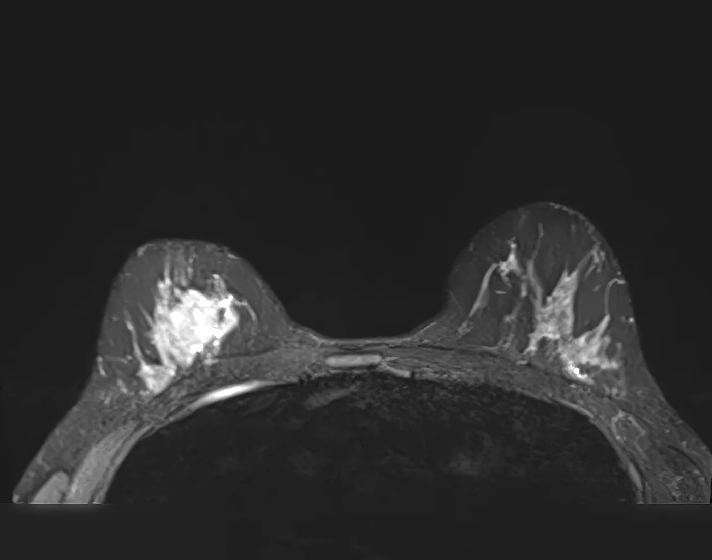
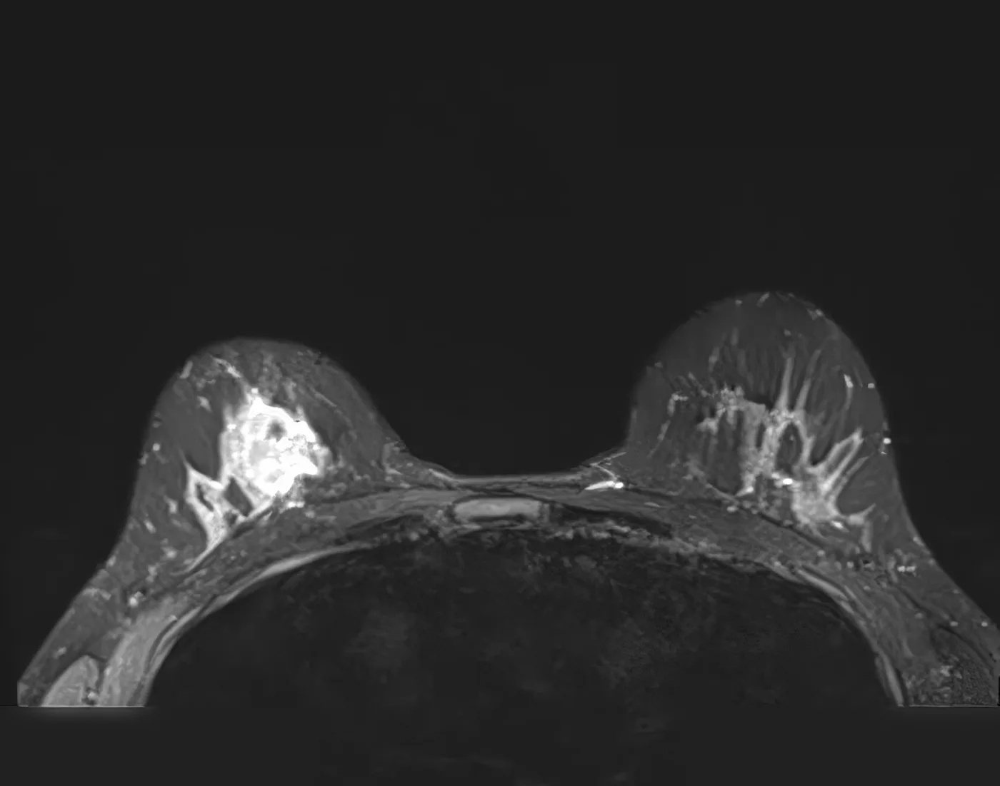
November 2024 · MRI (Month −2)INTERVAL PROGRESSION. Heterogeneously enhancing mass in the central right breast, at least 4.5 x 3.5 x 6.3 cm, significantly larger than prior. Multiple adjacent enhancing satellite lesions surrounding the dominant mass, new from prior exams. Enhancement extended posterosuperiorly and abutted underlying musculature without definitive muscle invasion. Left breast: no suspicious findings, no abnormal axillary nodes. BIRADS 6.
January 2025 · Session #1 · Day 0US-guided PEF ablation, 2 cycles, with intratumoral immunotherapy to the right breast mass, under ultrasound guidance with real-time visualisation of needle placement and ablation zone.
February 2025 · Session #2 · Day 37US-guided PEF ablation, 2 cycles, with intratumoral immunotherapy to the right breast mass.
May 2025 · Session #3 · Month 4US-guided PEF ablation, 1 cycle, with intratumoral immunotherapy to the right breast mass.
Total delivered5 PEF cycles plus intratumoral immunotherapy across three sessions.
November 2025 · Month 10 · Breast MRIPERSISTENT VIABLE TUMOUR. Interval increase in nodular soft tissue enhancement, especially lateral to the ablation zone, 2.2 x 1.4 cm, previously 1.8 x 1.1 cm. Overall disease extent 3.4 x 5.1 x 2.8 cm, extending from central to upper central and upper inner quadrants. Anterior non-mass enhancement, 1.8 cm, remained stable. Left breast: no suspicious enhancement, no enlarged nodes. IMPRESSION: persistent viable tumour in the right breast with slight increase in size compared with the Month 7 MRI. BIRADS 6. Surgical consultation recommended.
January 2026 · Month 12 · Intraoperative core needle biopsyFive tissue cylinders, 10x1 mm to 17x1 mm, light yellow rubbery tissue with few yellow soft areas. Microscopic: medium-sized mammary duct with increased epithelial layers and focal solid pattern, cells with medium-sized oval monotonous nuclei with fine chromatin, no mitosis; few bilayer lobules without nuclear atypia or mitosis adjacent to microcalcification; hypocellular collagenous stroma, mature adipose tissue and foamy macrophages. FINAL DIAGNOSIS: atypical ductal hyperplasia (ADH); chronic mastitis; NEGATIVE FOR NEOPLASTIC CELLS / MALIGNANCY.
Recent · PET/CT (per the clinic) Compared with the PET/CT at about Month 7: marked reduction of overall tumor burden and metabolic activity in the breast; a small residual focus remains at and around the marker clip from an earlier biopsy. Planned next: image-guided biopsy with focal ablation.
Imaging from the record
PET/CT · Baseline, before treatment
April 2024 · PET/CT · Baseline, before treatment
MRI · Before treatment · Month −2.5
November 2024 · MRI · Before treatment · Month −2.5
MRI · Before treatment · Month −2.5
November 2024 · MRI · Before treatment · Month −2.5
Ultrasound guidance · Session 1 · Day 0
January 2025 · Ultrasound guidance · Session 1 · Day 0
Ultrasound guidance · Session 1 · Day 0
January 2025 · Ultrasound guidance · Session 1 · Day 0
Ultrasound guidance · Session 1 · Day 0
January 2025 · Ultrasound guidance · Session 1 · Day 0
Ultrasound guidance · Session 2 · Day 37
February 2025 · Ultrasound guidance · Session 2 · Day 37
Ultrasound guidance · Session 2 · Day 37
February 2025 · Ultrasound guidance · Session 2 · Day 37
Ultrasound guidance · Session 2 · Day 37
February 2025 · Ultrasound guidance · Session 2 · Day 37
Ultrasound guidance · Session 3 · Month 4
May 2025 · Ultrasound guidance · Session 3 · Month 4
Ultrasound guidance · Session 3 · Month 4
May 2025 · Ultrasound guidance · Session 3 · Month 4
Ultrasound guidance · Session 3 · Month 4
May 2025 · Ultrasound guidance · Session 3 · Month 4
Is this for you?

Where this fits, stage by stage

Where your cancer is decides where we fit. Find the card closest to what you have been told. If you are unsure, send your records: a physician decides case by case, from your actual records.

Early (stage 0–I)

A small tumor, caught early. In the United States, freezing on its own is authorized with hormone therapy for women 70 or older with certain low-risk tumors. For some younger patients we may add immune drugs inside the tumor, decided case by case.

Locally advanced (II–III)

A larger tumor, or spread to nearby lymph nodes, but not beyond. Standard care is chemotherapy, sometimes with immunotherapy, then surgery and radiation. We may add immune drugs inside the tumor alongside that care.

Metastatic (stage IV)

Cancer has spread widely and the standard drugs have stopped working. We treat a tumor directly, to bring fresh immune cells to where the cancer is.

Oligometastatic

Cancer has spread, but only to a few places. Each spot can be treated directly while the immune system is engaged, aiming at a longer stretch without the disease growing.

You may be a candidate if:

Usually a tumor a needle can reach safely, plus one of these:

  • Your cancer is early and low-risk, and you want to avoid surgery
  • It is larger or in nearby lymph nodes and you are having standard treatment
  • It has spread to only a few places
  • The standard drugs have stopped working
  • You want to add immune treatment to the plan you are on
  • You are looking for options beyond your home country

It may not be suitable if:

  • You have inflammatory breast cancer that needs drug treatment first
  • The cancer is scattered too finely for a needle to target
  • You have a severe infection right now
  • You have a bleeding disorder that cannot be corrected

Each case is individually evaluated by the Williams Cancer Institute team.

Early (stage 0–I). Standard care: for women aged 70 or older with qualifying low-risk ER-positive tumors, ProSense cryoablation is authorized in the United States, together with endocrine therapy. Added here, under physician review: for selected younger patients, PEF or cryoablation with intratumoral immune priming.

Locally advanced (II–III). Standard care: neoadjuvant chemotherapy with or without immunotherapy, then surgery and radiation. Added here, under physician review: intratumoral immune priming alongside that care.

Oligometastatic. Disease confined to a few sites. Added here: local control of each site with immune activation, with the aim of a longer interval without progression than systemic therapy alone.

Metastatic (stage IV). Standard care: sequences of chemotherapy, checkpoint inhibitors, antibody–drug conjugates (Enhertu, Trodelvy), CDK4/6 inhibitors or PARP inhibitors. Added here, after progression on those: direct treatment of a tumor to engage fresh effector cells at the site of the antigen. WCI's rationale is that repeated IV immunotherapy exhausts T cells and that intratumoral delivery engages fresh ones.

WCI's stated candidacy criteria. Early-stage low-risk disease seeking a minimally invasive alternative to surgery · Locally advanced disease with a primary tumor target · Metastatic triple-negative, HER2-positive or hormone-receptor-positive disease progressing on standard sequences · Oligometastatic disease with limited progressing sites · Seeking systemic immune activation in addition to local control.

WCI's stated exclusions. Diffuse miliary metastatic pattern without a focal target · Active severe infection · Uncorrectable bleeding disorders · Very poor performance status (ECOG 3 or worse) · Inflammatory breast cancer requiring systemic therapy first. Each case is individually evaluated by the Williams Cancer Institute team.

How it works

Your Admissions Process, in Five Steps

After completing our admissions form, one of our Patient Consultants will contact you to explain the process and guide you through the next steps. Our admissions process is free of charge, including the review of your medical information and, if our medical team determines that you are a candidate, a video consultation. Treatments are generally not covered by health insurance, and WCI does not bill insurance companies.

  1. 01Free

    Initial Contact

    One of our Patient Consultants will contact you to explain the admissions process, answer your general questions, and guide you through the next steps.

  2. 02Free

    Medical Records and Imaging

    Your Patient Consultant will guide you on the required medical records and imaging studies, as well as how to submit them so our medical team can evaluate your case.

  3. 03Free

    Tumor Board Review

    Once the necessary information has been received, our medical team will review your case to determine whether you may be a candidate for our treatments. If necessary, the medical team may request additional studies or information before making a decision.

  4. 04Free

    Video Consultation

    If our medical team determines that you are a candidate, we will coordinate a complimentary Zoom consultation with one of our physicians. During this consultation, you will be able to discuss your case, address your medical questions, and learn about the treatment approach indicated for you.

  5. 05Self-pay treatment

    Treatment Plan and Next Steps

    Following your medical consultation, you will receive a personalized treatment plan that includes the corresponding costs. If you decide to proceed, the next steps will be coordinated to schedule your treatment.

    Most treatments take place in Los Cabos, Mexico; in some cases our medical team may schedule a procedure in Mexico City. Rebyota is given only at our Beverly Hills, California location.

For step 05

What to expect

Your physician confirms the details for your case.

Procedure
About an hour. Most people go home the same day.
Anesthesia or a stay
Conscious sedation is the predominant anesthesia used. It is rare that procedures require general anesthesia or an overnight stay. Your physician tells you which.
Number of treatments
Determined during the treatment plan design.
First check-up
6 to 8 weeks after the last treatment: a scan, and blood tests for your cancer type.
Your oncologist
Kept informed throughout.
Risks

As with any needle procedure, there can be bruising or bleeding at the site. Your physician goes through the risks for your case before anything is decided.

Records to send. Mammogram and breast ultrasound · Breast MRI, if done · Most recent PET/CT or CT · Biopsy pathology report · ER, PR, HER2 and Ki-67 results · BRCA, PD-L1 or TROP-2 results, if tested · A list of treatments so far, with dates. The physician confirms the list for each case.

Setting. Most procedures are outpatient under conscious sedation; general anesthesia or an overnight stay is rarely required.

Follow-up. First check-up at 6 to 8 weeks with a scan and the blood tests used for this cancer. The number of treatments is decided with the patient's physician.

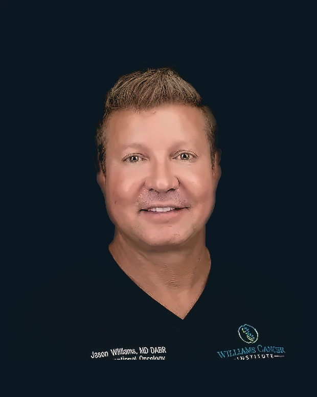
Dr. Jason R. Williams
Jason R. Williams, MD, DABRInterventional oncology
Dr. Williams holding the 2019 Vince Lombardi Cancer Foundation Leaders For A Cure award
Leaders For A Cure2019 · Vince Lombardi Cancer Foundation
The physician who decides every case

Dr. Jason R. Williams

MD, DABR · Director of Interventional Oncology and Immunotherapy

Dr. Jason Williams is a board-certified radiologist who has spent his career putting needles into tumors under imaging. His first patent, in 2003, covered ablation combined with immunotherapy injected into the tumor. A physician since 2000, he opened his first ablation center in 2004 and completed his residency in 2007. He also co-founded Syncromune, whose separate investigational drug SV-102 holds FDA Fast Track status. His tumor board reviews every case.

The team

Meet Our Medical Team

Tap a photo to read their background.
{{ teamSel.r }}

{{ teamSel.n }}

{{ para }}

Where treatment happens

Our Treatment Facilities in Los Cabos

Most of our treatments are performed in Los Cabos, Mexico, in a private hospital setting.

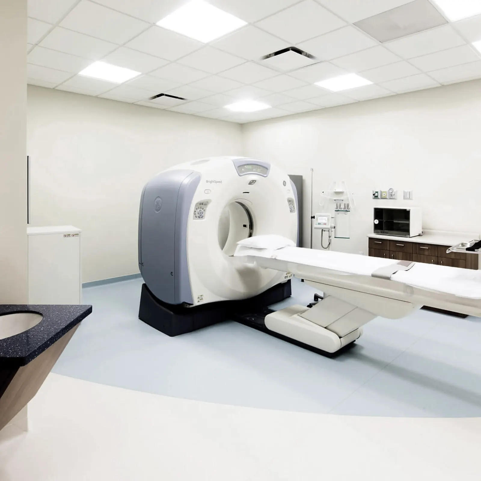
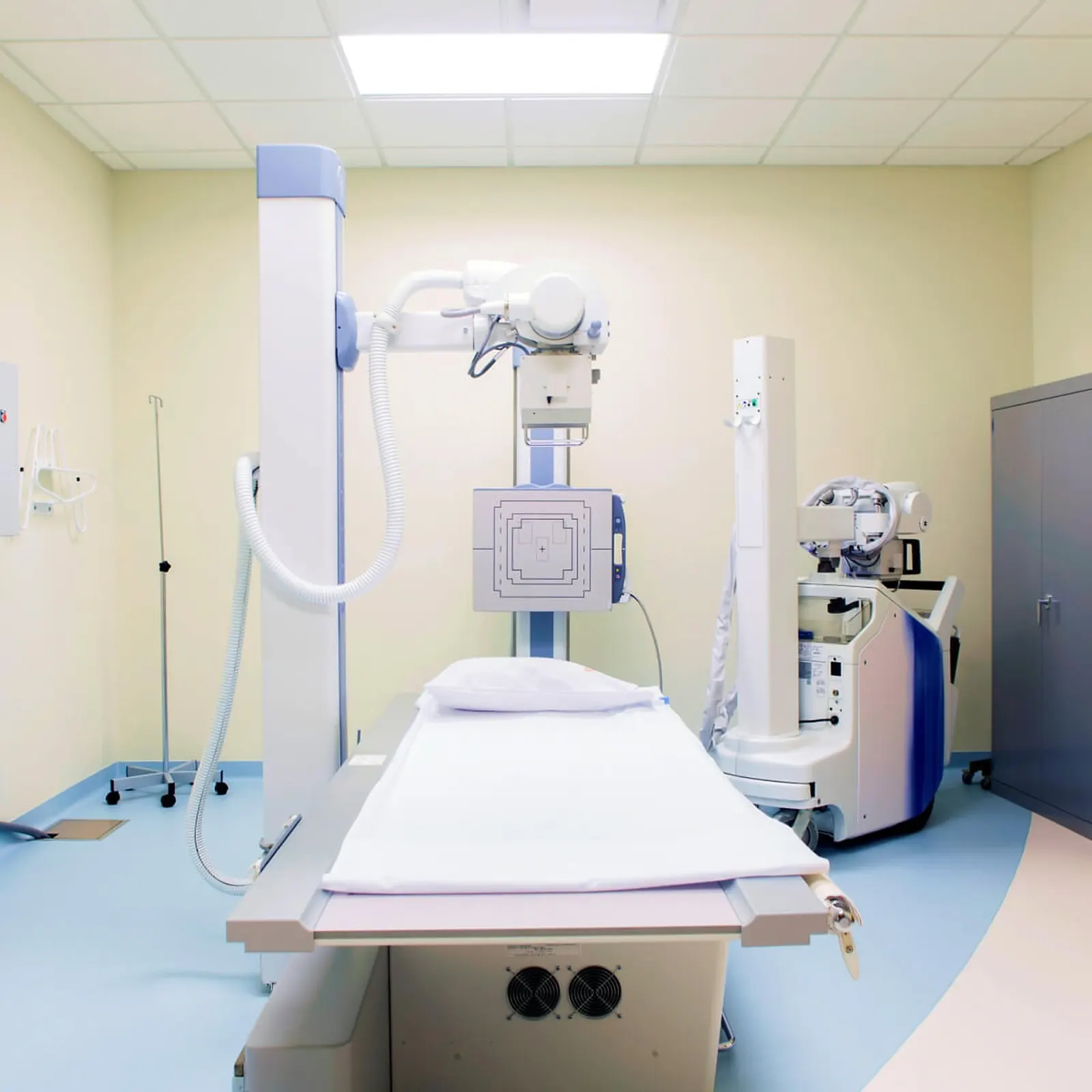
  • Imaging on site, including CT, ultrasound, MRI and digital radiology, for image-guided procedures.
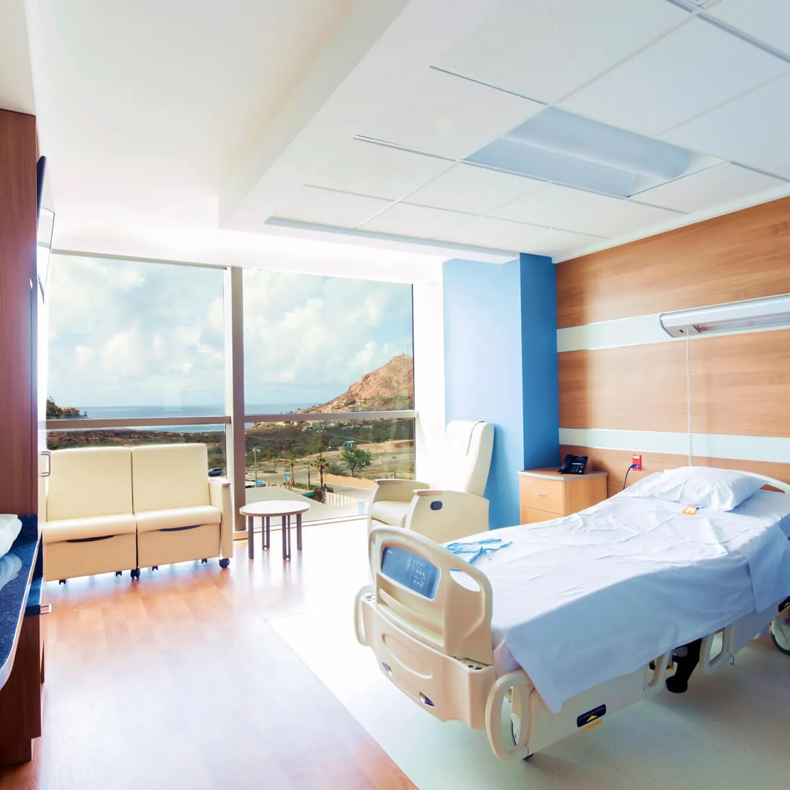
  • Private patient rooms. When an overnight stay is necessary, nursing care is available on the inpatient floor.
  • Located approximately 15 minutes from Los Cabos International Airport (SJD).

Other treatment locations. In some cases our medical team may schedule a procedure in Mexico City. Rebyota is given only at our Beverly Hills, California location.

Aerial view of the Los Cabos marina at sunset
Los Cabos
Waiting lounge overlooking the Sea of Cortez
Patient lounge
Private patient room with ocean view
Private room
CT procedure suite
Procedure suite
Digital radiology room
Radiology
Patient accounts

In their own words

Accounts Williams Cancer Institute publishes, shared with each patient's permission. They span different cancers. Individual results vary and are not guaranteed.

Videos

Breast Cancer on video

Selected Breast Cancer videos from the Williams Cancer Institute channel. Patient stories are shared with each patient's permission. Individual results vary.
The full Breast Cancer playlist on the Williams Cancer Institute YouTube channel.
Next step

Start Your Admissions Process

Complete our admissions form to get started. One of our Patient Consultants will contact you to explain the process and guide you through the next steps, including the medical information and imaging studies required for our medical team to evaluate your case.

info@williamscancerinstitute.com

Scan image not available in this build{{ zoomCap }}
{{ zoomCapShown }}